

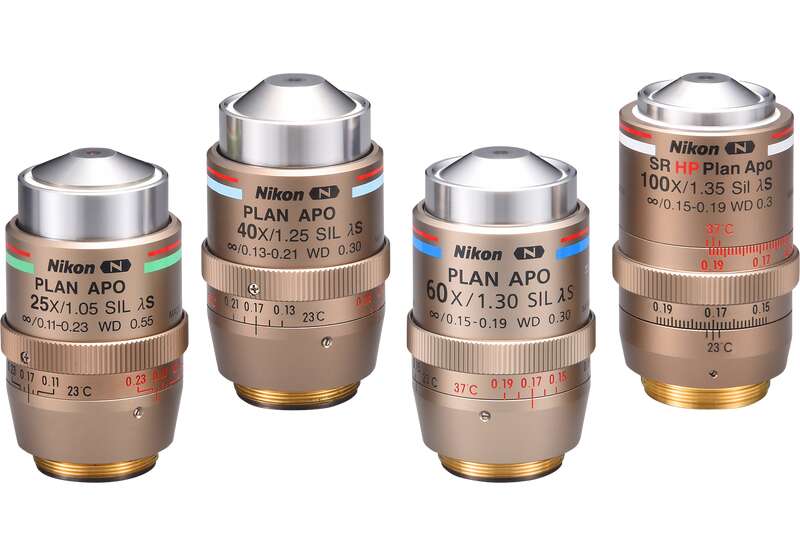
Overview & Features
Perfect for imaging 3D cell cultures and thick tissue samples. Silicone lenses enable visualization of cellular dynamics clearly and brightly when imaging at depth.
Research applications in neuroscience and cell biology using brain tissue, spheroids, organoids, and 3D cultures continue to push the limits of imaging in thick specimens. The need to image deep into such samples has never been more apparent. Nikon’s silicone immersion objectives enable clear observation with high signal-to-noise deep into living tissue.
The silicone immersion lens series features wide fields of view, high resolution, and evaporation-resistant oil, facilitating observations with ease.
With the addition of the new 60X objective, which employs newly developed glass for enhanced chromatic aberration correction, quantitative imaging in thick living specimens has never been more attainable.
Features
Key Features
Efficiently switch magnification for macro-to-micro imaging without changing the immersion medium
SIL25X

Zoom 1X, Pixel 0.69 μm

Zoom 5X, Pixel 0.14 μm
SIL40X

Zoom 1X, Pixel 0.43 μm

Zoom 4X, Pixel 0.11 μm
SIL60X

Zoom 3X, Pixel 0.10μm

Zoom 1X, Pixel 0.29 μm
Small intestine enteroid (Cadherin: Alexa Fluor® 555, Nuclear: DAPI)
Images courtesy of: Dr. Yuki Yokoi, Dr. Kiminori Nakamura, and Dr. Tokiyoshi Ayabe
Innate Immunity Laboratory, Department of Cell Biological Science, Faculty of Advanced Life Science
Graduate School of Life Science, Hokkaido University
SIL100X

Zoom 1.6X, Pixel 0.11 μm

Zoom 1X, Pixel 0.17 μm
Make high-resolution deep observations of thick specimens such as 3D cell cultures, organoids, and tissues


Small intestine enteroid (Cadherin: Alexa Fluor® 555, Nuclear: DAPI)
Images courtesy of: Dr. Yuki Yokoi, Dr. Kiminori Nakamura, and Dr. Tokiyoshi Ayabe
Innate Immunity Laboratory, Department of Cell Biological Science, Faculty of Advanced Life Science
Graduate School of Life Science, Hokkaido University Science, Hokkaido University
Observe live samples over long periods of time
It is also best suited for long time-lapse imaging of live cells without the immersion medium evaporating because silicone oil has low volatility even at 37°C.


Time-lapse imaging of enteroid (25X)
Images courtesy of: Dr. Yuki Yokoi, Dr. Kiminori Nakamura, and Dr. Tokiyoshi Ayabe
Innate Immunity Laboratory, Department of Cell Biological Science, Faculty of Advanced Life Science
Graduate School of Life Science, Hokkaido University
By using the Denoise.ai in conjunction with the AX confocal microscope, you can acquire even sharper images.


Newly developed Short-wavelength Refractive (SR) glass
The new silicone immersion 60X objective employs high- and specialized-dispersion glass that was independently developed by Nikon and possesses extra-low dispersion properties.
By refracting short-wavelength light to a higher degree, it is possible to collect a wider range of wavelengths, resulting in significantly enhanced chromatic aberration correction. In addition to axial chromatic aberrations, lateral chromatic aberrations can also be corrected.
Using only normal lens

With SR Lens

*The photo is a sample image
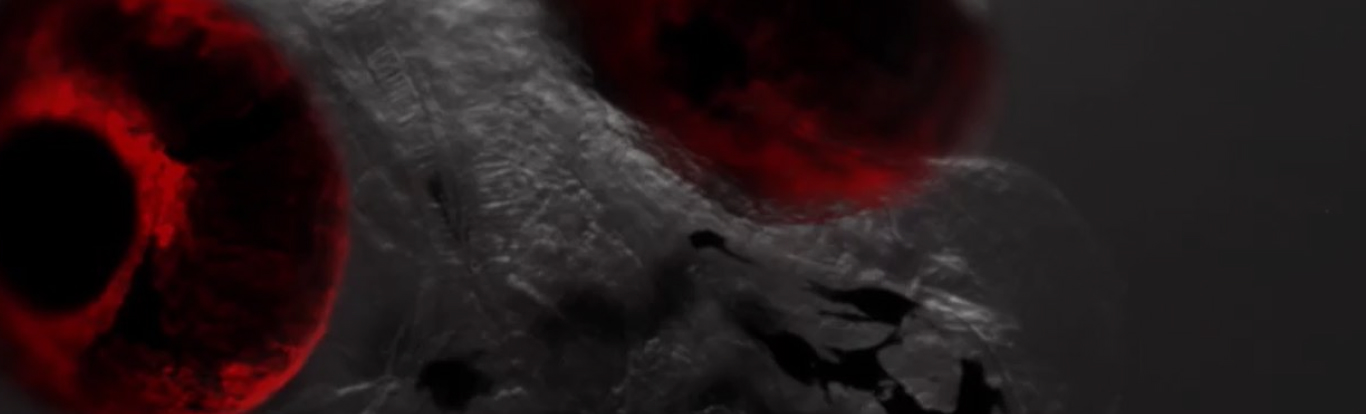
Useful for a variety of scientific disciplines such as immunology, developmental biology, neuroscience, drug discovery and regenerative medicine.
Images courtesy of: Lin Daniel, PhD. SunJin Lab Co.

Images courtesy of: Professor Masaru Ishii, Department of Immunology and Cell Biology, Graduate School of Medicine, Osaka University


Image courtesy of: Dr. Hidenori Akutsu and Dr. Tomoyuki Kawasaki of the Center for Regenerative Medicine, National Center for Child Health and Development

Image courtesy of: Dr. Yoshiteru Kai, Reproductive Medicine Research Center, YAMASHITA SHONAN YUME CLINIC

Specifications
| Model | Dimensions | Transmittance | NA | W.D. (mm) | Cover glass thickness | Correction ring | Observation |
|---|---|---|---|---|---|---|---|
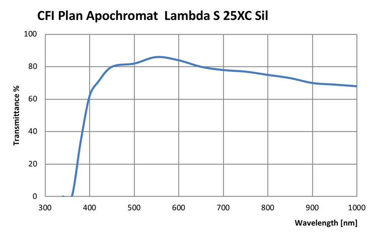
| CFI Plan Apochromat Lambda S 25XC Sil | Diagram | Graph | 1.05 | 0.55* | 0.11 – 0.23 | ∨ | BF, DF, DIC, POL, FL (visible light, UV, NIR) |
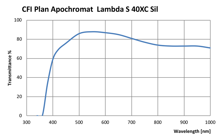
| CFI Plan Apochromat Lambda S 40XC Sil | Diagram | Graph | 1.25 | 0.3* | 0.13 – 0.21 (23˚C) 0.15 – 0.23 (37˚C) | ∨ | BF, DF, DIC, POL, FL (visible light, UV, NIR) |
| CFI Plan Apochromat Lambda S 60XC Sil | Diagram | Graph | 1.30 | 0.3* | 0.15 – 0.19 | ∨ | BF, DIC, POL, FL (visible light, UV, NIR) |
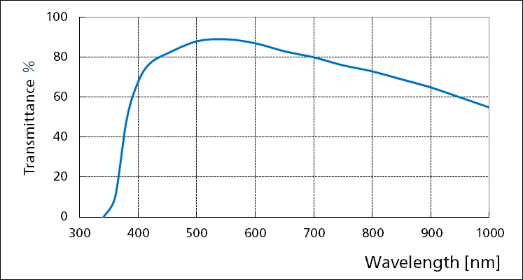
| CFI SR HP Plan Apochromat Lambda S 100X Sil | Diagram | Graph | 1.35 | 0.30* (0.31 – 0.29) (23˚C) 0.29* (0.30 – 0.28) (37˚C) | 0.15 – 0.19 | ∨ | BF, DIC, POL, FL (visible light, UV, NIR) |
BF: Brightfield
DF: Darkfield
DIC: Differential Interference Contrast
POL: Simple polarizing
FL: Fluorescence
* With cover glass thickness of 0.17 mm