

Products for Drug Discovery
Inverted Microscope Systems
ECLIPSE Ti2-E / Ti2-LAPP: Synchronous operation of the Ti2-E inverted microscope with Ti2-LAPP illuminator modules and other hardware devices allows for complex multidimensional image acquisitions. Additionally, the Ti2-E features the Perfect Focus System 4 (PFS4) focus-locking system, providing the highest level of stability for extended imaging.
ECLIPSE Ji: The Ji is a digital microscope developed to support drug discovery research. Pre-defined assays are available to automatically perform numerous routine experiments, such as counting cells, measuring fluorescence expression efficiency, and measuring apoptosis. The instrument takes care of troublesome focus and exposure adjustments and supports the analysis of experimental results by displaying results per cell and per plate in an intuitive graphical interface.
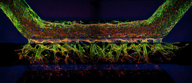
 3D image stack of an angiogenesis model cultured using the OrganoPlate® system (MIMETAS).
High Content Imaging
BioPipeline LIVE / PLATE: Nikon offers the BioPipeline LIVE and BioPipeline PLATE systems for high throughput and high content imaging. Both systems are based on our ECLIPSE Ti2-E inverted microscope and feature a robotic arm for automatic exchange and imaging of up to 44 vessels, including well plates. The BioPipeline LIVE additionally includes full incubation coverage for long-term live cell imaging-based assays.
Confocal Imaging
AX / AX R: The AX / AX R point-scanning confocal systems are capable of imaging over a wide 25 mm field of view with up to 8192 x 8192-pixel resolution. The AX R is equipped with a resonant scanner capable of very high-speed imaging (2048 x 512 pixels, 1024 x 512 pixels: 30 frames per second), supporting high-throughput screening, live cell imaging, and more.
CSU Series Spinning Disk Confocal Systems: The CSU-W1, and CSU-W1 SoRa spinning disk confocal from Yokogawa can all be configured on the BioPipeline PLATE and LIVE systems, providing flexible, live cell friendly confocal options for high content imaging.
Multiphoton Imaging
AX R MP: The AX R MP is Nikon’s latest multiphoton system, providing deep imaging up to 1.4 mm into the sample using 1300 nm excitation light, making it ideal for late in vivo and intravital imaging. Two dedicated upright microscope stands are available for use with the AX R MP – the single stand for wide samples and the gate stand for deep ones.
Software and AI
NIS-Elements Software: NIS-Elements is Nikon’s flagship, integrated software solution for microscope operation, image analysis, and visualization. NIS-Elements HC is optimized for high-throughput and high-content imaging applications. Heatmaps, sample images, binary masks, assay results, and more can be centrally managed for rapid filtering and analysis. Additionally, the NIS.ai deep learning-based software modules can be incorporated to harness the power of artificial intelligence (AI) for analyses such as image segmentation.
●: included, ⚬: option
| Base System | Modules | ||||||
|---|---|---|---|---|---|---|---|
| ECLIPSE Ti2-E Inverted Microscope (Widefield Imaging) |
ECLIPSE Ji Digital Inverted |
AX R Resonant Confocal System |
Ti2-LAPP E-TIRF Illuminator |
AX R MP Multiphoton Microscope System |
CSU-W1 Spinning Disk Confocal Scanner |
CSU-W1 SoRa Spinning Disk Super-Resolution System |
|
| Relative Imaging Depth Limit | ~ 5 μm ~ 15 – 25 μm (with deconvolution) |
~ 5 μm ~ 15 – 25 μm (with deconvolution) |
~ 100 – 500 μm | ~ 100 – 300 nm | ~ 500 μm – 1.5 mm | ~ 50 – 100 μm | ~ 50 – 100 μm |
| Supports Video Rate Imaging | ●*1 | ●*1 | ●*2 | ●*1 | ●*2 | ●*3 | ●*3 |
| Field of View | 25 mm diagonal (circular) | 25 mm diagonal (square) | 25 mm diagonal (square) | ~ 10 mm diagonal (circular) | 22 mm diagonal (square) | 17 x 16 mm (rectangular) | 17 x 16 mm (rectangular) |
Supported Imaging Modalities
| ECLIPSE Ti2-E |
ECLIPSE Ji |
AX R | Ti2-LAPP | AX R MP | CSU-W1 | CSU-W1 SoRa |
|
|---|---|---|---|---|---|---|---|
| Brightfield | ● | ● | |||||
| Confocal | Point-Scanning | Point-Scanning | Spinning Disk | Spinning Disk | |||
| Darkfield | ● | ||||||
| DIC | ● | ||||||
| Nikon Advanced Modulation Contrast (NAMC) | ● | ||||||
| Phase Contrast | ● | ||||||
| Super-Resolution | ISM with NSPARC detector | Optical Pixel Reassignment | |||||
| TIRF | ● | ||||||
| Volume Contrast | ● | ● | |||||
| Widefield Fluorescence | ● | ● | ● |
Compatible Microscope Stands
| ECLIPSE Ti2-E | ECLIPSE Ji | AX R | Ti2-LAPP | AX R MP | CSU-W1 | CSU-W1 SoRa | |
|---|---|---|---|---|---|---|---|
| ECLIPSE Ti2 Series Inverted | Ti2-E | Ti2-E | Ti2-E | ● | ● | ||
| ECLIPSE Ji Digital inverted | ● |
||||||
| ECLIPSE Ni Series Upright | Ni-E | ||||||
| ECLIPSE FN1 Upright | ● | ||||||
| AX-FNGP Upright | ● |
||||||
| AX-FNSP Upright | ● |
High Content Imaging Compatibility
| ECLIPSE Ti2-E | ECLIPSE Ji |
AX R | Ti2-LAPP | AX R MP | CSU-W1 | CSU-W1 SoRa | |
|---|---|---|---|---|---|---|---|
| BioPipeline LIVE | ● | ● | ● | ● | ● | ||
| BioPipeline PLATE | ● | ● | ● | ● | ● |
*1 Limited by camera system
*2 30 FPS with 512 x 512 scan
*3 Limited by camera system and disk rotation speed
Related Literature

Precision Imaging in Complex Tissue Structures
Tháng 6 2025
AI-powered “Sample Navigation” simplifies image acquisition! Improved reproducibility and streamlined information sharing
Tháng 6 2025
Confocal imaging of immune cells in a vasculature network using the HUMIMIC Chip
Tháng 6 2025
3D image analysis reveals drug-induced cellular senescence and increased nucleolus volume: Efficient imaging methods to maximize data acquisition
Tháng 6 2025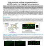
High-sensitivity confocal microscope detector NSPARC enables live imaging of autophagosomes.
Tháng 6 2025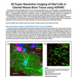
3D Super-Resolution Imaging of Glial Cells in Cleared Mouse Brain Tissue using NSPARC
Tháng 6 2025
Image-based analysis of intracellular delivery of DNA/RNA therapeutics using NSPARC
Tháng 6 2025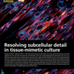
Resolving subcellular detail in tissue-mimetic culture
Tháng 6 2025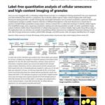
Label-free quantitative analysis of cellular senescence and high-content imaging of granules
Tháng 6 2025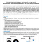
Episcopic brightfield imaging of neuronal cells on high-density microelectrode arrays enables prediction of cell region through AI learning
Tháng 6 2025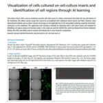
Visualization of cells cultured on cell-culture inserts and identification of cell regions through AI learning
Tháng 6 2025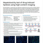
Hepatotoxicity test of drug-induced lipidosis using high-content imaging
Tháng 6 2025
Validation of control cells in phenotypic screening and selection of analysis methods according to cell morphology
Tháng 6 2025
Cell cycle analysis of keratinocytes and label-free cell counting of mitotic cells using Volume Contrast
Tháng 6 2025
Confocal Imaging of CAR-T Cell Dynamics Using an Organ-on-a-chip Platform
Tháng 6 2025
Cell Proliferation Assay and Optimization of HCA Assay, Using Label-Free Live Cell Imaging
Tháng 6 2025
Low Phototoxicity Long-Term Live Cell Apoptosis Assay Using Label-free Live Cell Imaging
Tháng 6 2025
Quantitative analysis of mitochondrial morphology over time using AI
Tháng 6 2025
Selecting the Right Objectives – Bright, Sharp Imaging of Structures down to Deep Areas
Tháng 6 2025
3D Imaging of Intestinal Organoid
Tháng 6 2025
Quantitative 3D Imaging of Living Organs-on-Chips with a High-Speed Point-Scanning Confocal System
Tháng 6 2025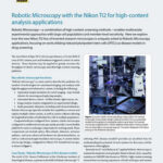
Robotic Microscopy with the Nikon Ti2 for High-Content Analysis Applications
Tháng 6 2025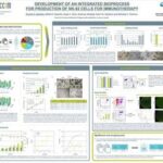
Development of an Integrated Bioprocess for Production of NK-92 Cells for Immunotherapy
Tháng 6 2025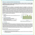
Antitumor Lymphocyte Kinetic Cytotoxicity Assay (Collaboration with CCRM)
Tháng 6 2025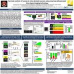
Survival Analysis of Human In Vitro-derived Neurons Using New Live Cell Extended Time-lapse Imaging Technology
Tháng 6 2025
3D Confocal Imaging of Thick Samples Using Z Intensity Correction
Tháng 6 2025Discussion of Drug Discovery
Selecting a Microscope System for your Drug Discovery Model
Model systems for drug discovery run just about the entire range of possibility, from in vitro adherent cell cultures to whole model organisms and almost everything in between. Complex 3D cell culture models such as spheroids, organoids, and organs-on-chips can be composed of multiple cell types to better recapitulate various physiological characteristics lost in traditional adherent cell cultures, which are the traditional standard for high throughput screening. Additionally, organoids can even be cultured from autologous (patient-derived) cells for developing precision medicines.
Widefield fluorescence imaging is an appropriate choice for relatively flat samples, such as adherent cells cultured in multi-well plates, the traditional standard for high throughput screening. It is fast, sensitive, and cost effective, but does not provide intrinsic optical sectioning – the ability to effectively image a single plane in a thick 3D specimen.
Physically larger 3D models, such as organoids and tissue, may require an imaging technique with optical sectioning capabilities to capture the features of interest without being overly compromised by out-of-focus blur. Confocal microscopy is the standard for such applications, Nikon offers the AX / AX R confocal microscopes, point-scanning systems which can be configured on the BioPipeline LIVE and BioPipeline PLATE systems for imaging discrete sections up to several hundred micrometers deep in various samples.
While confocal imaging is a suitable choice for optical sectioning at depth, sometimes it is insufficient. In vivo imaging in the presence of thick and scattering tissues often necessitates the use of multiphoton imaging, such as the Nikon AX R MP multiphoton microscope system, which uses multiphoton excitation with near-infrared to infrared light to minimize out-of-focus excitation and scattering.
Enhancing Drug Discovery Research using Artificial Intelligence
One of the great advantages of image-based assays is their rich information content. Until recently, however, relatively little of this information could be leveraged in a practical manner. However, new approaches based on artificial intelligence (AI), and particularly deep learning (DL) methods using artificial neural networks (ANNs), allow for deeper correlations between image features to be inferred and applied towards characterizing morphology and phenotype. Such analytical approaches have been referred to as “cell profiling” or “image-based profiling,” and represent a very active area of development.
In addition to profiling, DL can be used to help accelerate and strengthen image analyses in other ways. Nikon is committed to the design of reliable DL-based image analysis tools under the umbrella of NIS.ai – a series of software modules available for the NIS-Elements software. For example, the Segment.ai module can be trained to automatically segment difficult image features that are difficult to isolate using classical approaches.
Other NIS.ai modules of potential application for drug discovery work includes Convert.ai, which can be trained to predict image features from a fluorescence channel using only a brightfield or another transmitted light channel as a reference, which can help cut down on both cytotoxicity (from the fluorescent label) and phototoxicity (from the high intensity light used for fluorescence imaging). Relatedly, the Enhance.ai module can be trained to predict a higher signal-to-noise version of noisy image data. This allows for a reduction in illumination intensity, and thus phototoxicity.
High throughput widefield fluorescence imaging of larger model systems, such as organoids, can benefit from our Clarify.ai module, which is pre-trained to provide automatic blur removal from widefield fluorescence microscopy images. This allows for users to benefit from the speed of widefield fluorescence imaging, but with enhanced optical sectioning and without the need for a confocal system.
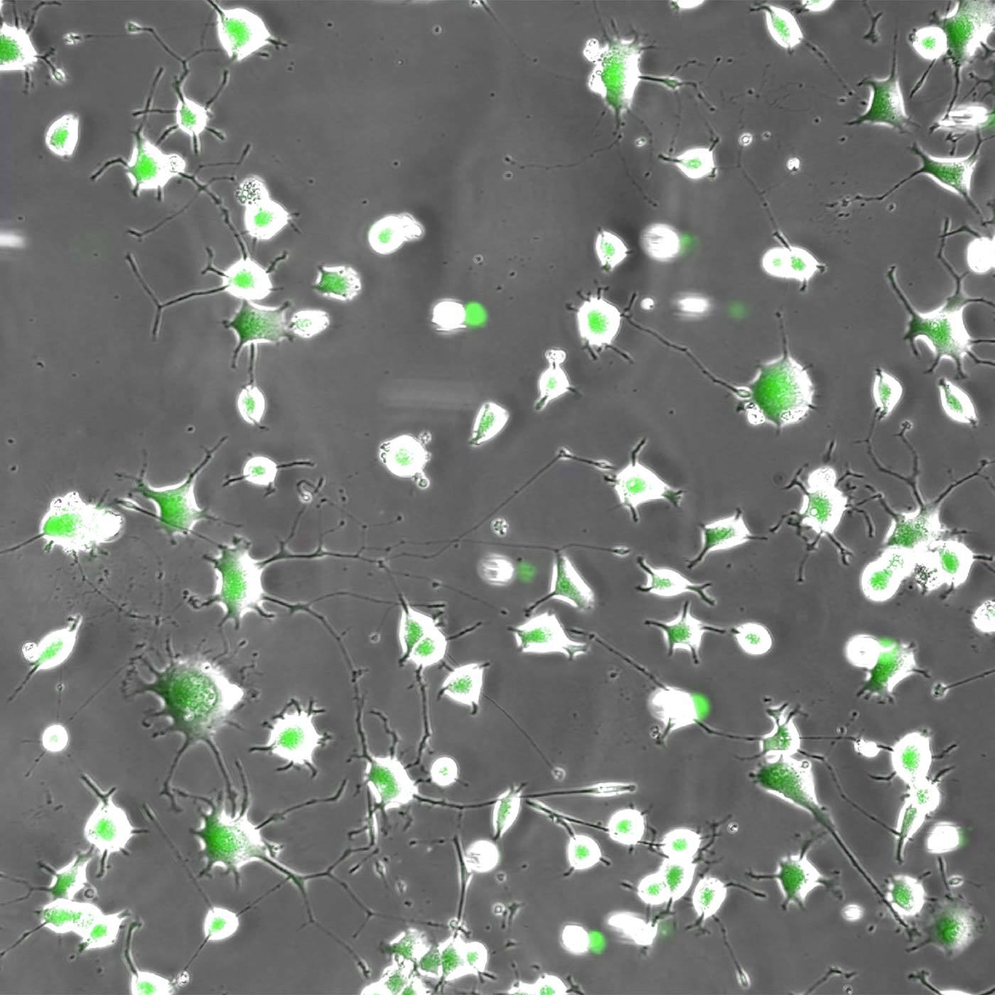 Original
Neurites in phase-contrast were not possible to define accurately by traditional thresholding. Segment.ai was trained on hand-traced neurites (human recognized) and learned how to trace neurites in subsequent images.
Nikon Contract Imaging Services for Pre-Clinical Drug Discovery Research
The Nikon BioImaging Labs provide contract research services to their local biotech and research communities, as well as remote services* for clients located elsewhere. With locations in Cambridge, MA, USA, Leiden, The Netherlands, and Shonan, Japan, the Nikon BioImaging Labs have significant experience working with customers in the biopharma research space and performing confocal microscopy of organoids and other 3D cell culture systems now used for drug discovery studies, including various commercially produced organ-on-chip models.

The Nikon BioImaging Labs are not only capable of data acquisition, but their full-service capabilities also extend to assay design, assay development, assay validation, cell culture, sample preparation, and data analysis. If you would like to learn more and see if services at one of the Nikon BioImaging Labs is right for you, please don’t hesitate to contact us to set up a free consultation.
* The service may vary depending on the facility. Please contact the Nikon BioImaging Lab near your location for details.
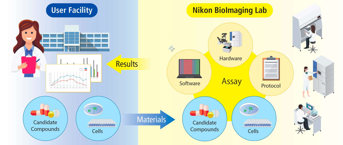
Nikon contract research services at a glance.