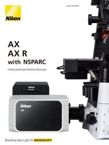

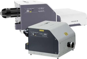
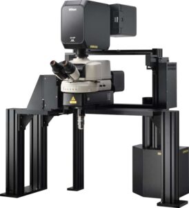
Confocal microscopy provides optical sectioning, the ability to observe discrete planes in 3D samples, by using one or more apertures to block out-of-focus light. Nikon offers both point-scanning confocal instruments, led by our AX / AX R confocal system, as well as spinning disk (field scanning) confocal systems from top manufacturers.
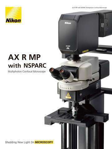
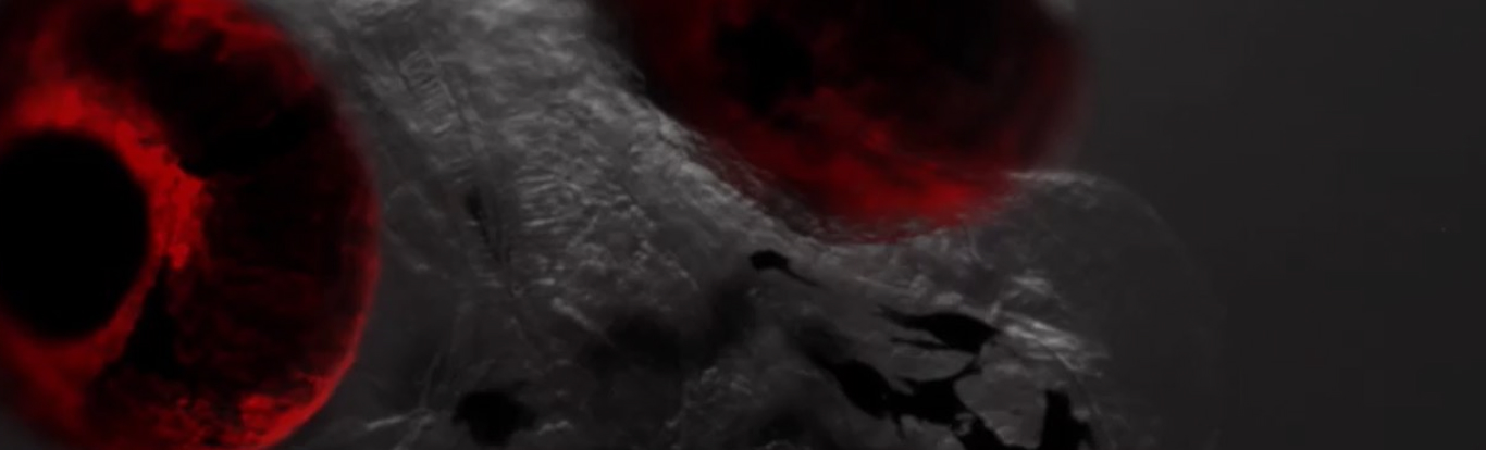
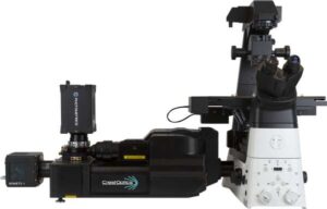
Multiphoton microscopy is preferred for deep imaging applications in thick specimens, including intravital imaging. Non-linear excitation restricts fluorescence to the laser focus and near-infrared illumination minimizes absorption and scattering. Nikon offers the AX R MP multiphoton system, available with microscope stand options optimized for large specimens.
Image scanning microscopy (ISM) is a super-resolution technique that takes advantage of structured detection of each point in a point-scanning system to improve both resolution and signal-to-noise (S/N), a great choice for low light imaging. Both the AX / AX R confocal and AX R MP multiphoton systems may be equipped with the Nikon Spatial Array Confocal (NSPARC) detector for ISM imaging.
Product Lineup
Related Literature
Feedback microscopy using Nikon AX confocal microscopy and JOBS software to bridge mm to µm structures
Tháng 6 2025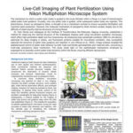
Live-Cell Imaging of Plant Fertilization Using Nikon Multiphoton Microscope System
Tháng 6 2025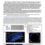
3D Imaging of Kidney Proximal Tubule Microphysiological Systems
Tháng 6 2025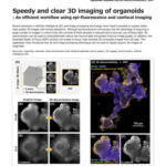
Speedy and clear 3D imaging of organoids
Tháng 6 2025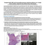
Imaging both H&E and immunofluorescence staining patterns on a single tissue section using the AX confocal laser microscope
Tháng 6 2025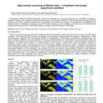
High content screening of lifetime data – A feedback microscopy experiment workflow
Tháng 6 2025
Precision Imaging in Complex Tissue Structures
Tháng 6 2025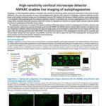
High-sensitivity confocal microscope detector NSPARC enables live imaging of autophagosomes.
Tháng 6 2025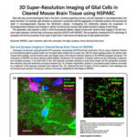
3D Super-Resolution Imaging of Glial Cells in Cleared Mouse Brain Tissue using NSPARC
Tháng 6 2025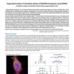
Image-based analysis of intracellular delivery of DNA/RNA therapeutics using NSPARC
Tháng 6 2025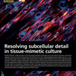
Resolving subcellular detail in tissue-mimetic culture
Tháng 6 2025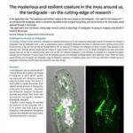
The mysterious and resilient creature in the moss around us, the tardigrade – on the cutting-edge of research –
Tháng 6 2025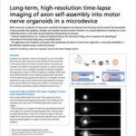
Long-term, high-resolution time-lapse imaging of axon self-assembly into motor nerve organoids in a microdevice
Tháng 6 2025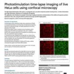
Photostimulation time-lapse imaging of live HeLa cells using confocal microscopy
Tháng 6 2025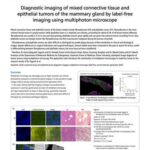
Diagnostic imaging of mixed connective tissue and epithelial tumors of the mammary gland by label-free imaging using multiphoton microscope
Tháng 6 2025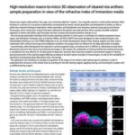
High-resolution macro-to-micro 3D observation of cleared rice anthers: sample preparation in view of the refractive index of immersion media
Tháng 6 2025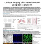
Confocal Imaging of in vitro BBB model using idenTx platform
Tháng 6 2025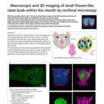
Macroscopic and 3D imaging of small flower-like taste buds within the mouth by confocal microscopy
Tháng 6 2025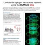
Confocal imaging of vasculature network using the HUMIMIC Chip
Tháng 6 2025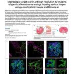
Macroscopic target search and high-resolution 3D imaging of gastric afferent nerve endings showing various shapes using a confocal microscope and Denoise.ai
Tháng 6 2025
Quantitative Analysis of Mitochondrial Microstructure by Super-Resolution Imaging
Tháng 6 2025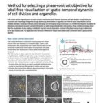
Method for selecting a phase-contrast objective for label-free visualization of spatio-temporal dynamics of cell division and organelles
Tháng 6 2025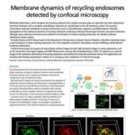
Membrane dynamics of recycling endosomes detected by confocal microscopy
Tháng 6 2025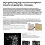
High-speed, deep, high-resolution multiphoton imaging using expansion microscopy
Tháng 6 2025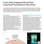
In vivo Deep Imaging of Mouse Brain using Novel Fluoropolymer Nanosheet
Tháng 6 2025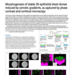
Morphogenesis of stable 3D epithelial sheet domes induced by osmotic gradients, as captured by phase contrast and confocal microscopy
Tháng 6 2025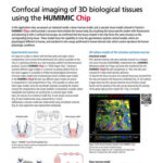
Confocal imaging of 3D biological tissues using the HUMIMIC Chip
Tháng 6 2025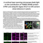
A confocal laser scanning microscope sheds light on the contribution of TGN/EE SNARE protein SYP61 and ubiquitin ligase ATL31 to C/N-nutrient stress tolerance in plants
Tháng 6 2025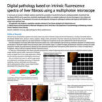
Digital pathology based on intrinsic fluorescence spectra of liver fibrosis using a multiphoton microscope
Tháng 6 2025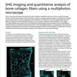
SHG imaging and quantitative analysis of bone collagen fibers using a multiphoton microscope
Tháng 6 2025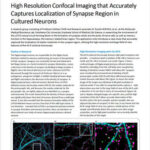
High Resolution Confocal Imaging that Accurately Captures Localization of Synapse Region in Cultured Neurons
Tháng 6 2025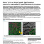
Macro to micro skeletal muscle fiber formation mechanism captured with large FOV confocal microscope
Tháng 6 2025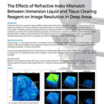
The Effects of Refractive Index Mismatch Between Immersion Liquid and Tissue Clearing Reagent on Image Resolution in Deep Areas
Tháng 6 2025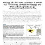
Ecology of a fossilized cockroach in amber was revealed by confocal microscopy and thin sectioning technology
Tháng 6 2025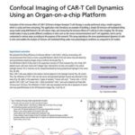
Confocal Imaging of CAR-T Cell Dynamics Using an Organ-on-a-chip Platform
Tháng 6 2025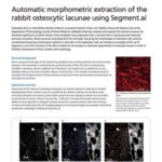
Automatic morphometric extraction of the rabbit osteocytic lacunae using Segment.ai
Tháng 6 2025
Morphogenesis imaging of mCherry-expressing Magnaporthe oryzae transformants with DIC and confocal microscopy
Tháng 6 2025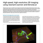
High-speed, high-resolution 3D imaging using resonant scanner and Denoise.ai
Tháng 6 2025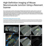
High-Definition Imaging of Mouse Neuromuscular Junction Using a Resonant Scanner
Tháng 6 2025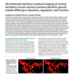
3D enhanced resolution confocal imaging of cortical excitatory mouse neurons uncovers dendritic spinule subsets differing in dynamics, regulation, and function
Tháng 6 2025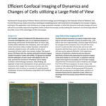
Efficient Confocal Imaging of Dynamics and Changes of Cells utilizing a Large Field of View
Tháng 6 2025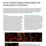
In vivo Confocal Imaging of Mouse Organs that Clearly Captures Fast Dynamics
Tháng 6 2025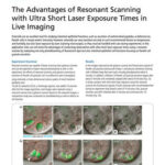
The Advantages of Resonant Scanning with Ultra Short Laser Exposure Times in Live Imaging
Tháng 6 2025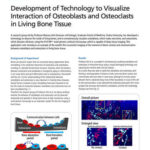
Development of Technology to Visualize Interaction of Osteoblasts and Osteoclasts in Living Bone Tissue
Tháng 6 2025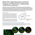
Wide FOV, High-Resolution Confocal Imaging of Podosomes in Osteoclasts ~ Macro and micro observation ~
Tháng 6 2025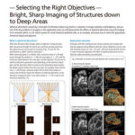
Selecting the Right Objectives – Bright, Sharp Imaging of Structures down to Deep Areas
Tháng 6 2025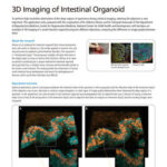
3D Imaging of Intestinal Organoid
Tháng 6 2025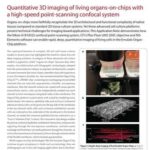
Quantitative 3D Imaging of Living Organs-on-Chips with a High-Speed Point-Scanning Confocal System
Tháng 6 2025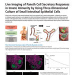
Live Imaging of Paneth Cell Secretory Responses in Innate Immunity by Using Three-Dimensional Culture of Small Intestinal Epithelial Cells
Tháng 6 2025
The Secret Inside Flowers – Imaging Inside Plants Using ClearSee Clearing Reagent
Tháng 6 2025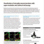
Visualization of microglia-neuron junctions with super-resolution and confocal microscopy
Tháng 6 2025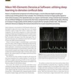
Nikon NIS-Elements Denoise.ai Software: utilizing deep learning to denoise confocal data
Tháng 6 2025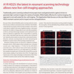
A1R HD25: the latest in resonant scanning technology allows new live-cell imaging approaches
Tháng 6 2025
Increasing Data Collection and Fidelity by Maximizing Confocal Field of View
Tháng 6 2025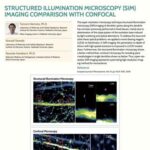
Structured Illumination Microscopy (SIM) Imaging Comparison with Confocal
Tháng 6 2025