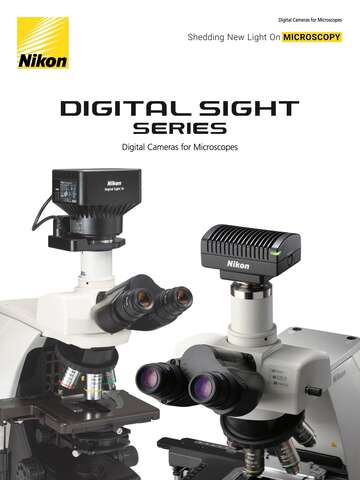

Overview & Features
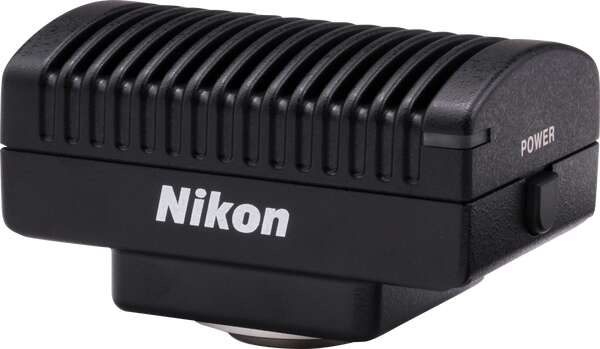
DS-Fi3 is a high-definition color microscope camera equipped with a 5.9 megapixel CMOS image sensor. Its high-speed data readout, superior color reproduction and high quantum efficiency are optimal for imaging in various observations, such as brightfield, DIC, phase contrast and fluorescence observation.
* DS-Fi3 is not for clinical diagnostic use.
Features
High definition imaging
DS-Fi3 is equipped with a 5.9 megapixel CMOS image sensor, which enables the capture of high-definition images of up to 2880 x 2048 pixels. With this new CMOS image sensor and high-speed data transfer via USB 3.0, DS-Fi3 enables fast focusing even in high-resolution imaging, and efficient image acquisition when using a wide range of illumination techniques.
High sensitivity and low noise
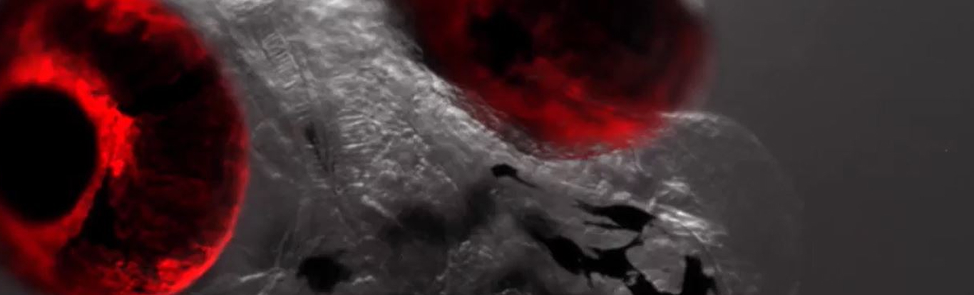
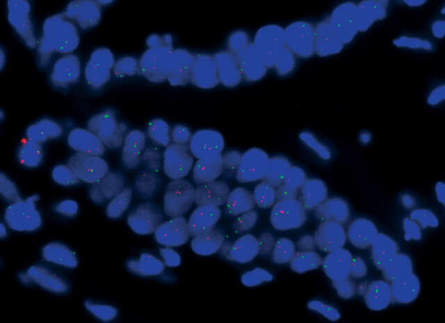
The DS-Fi3 has significantly higher quantum efficiency and lower readout noise than conventional models. It allows for the capture of brighter images with higher S/N ratios in fluorescence observation modes.

High-speed live display
Due to the high-speed data readout by the CMOS image sensor and high-speed USB3.0 data transfer, the DS-Fi3 displays live images of full 2880 x 2048-pixel images at 15 fps, and 1440 x 1024-pixel images at 30 fps. This enables smooth display during focusing and the selection of observation locations.
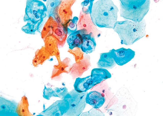
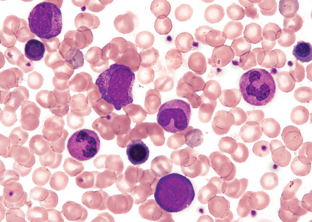
Excellent color reproducibility
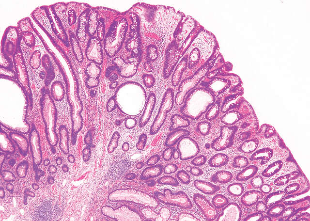
Nikon is well known for outstanding and lifelike color reproduction, and developing superior algorithms for creating results that look like the actual samples. These algorithms are used in all of the color cameras in the digital sight lineup.
Direct connection to a PC
The DS-Fi3 allows direct connection to a PC via a USB3.0 interface to control the camera and capture images using NIS-Elements imaging software.
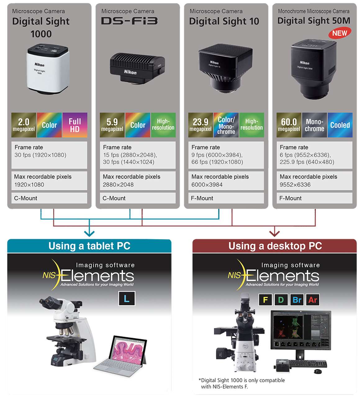
Different camera control options
Specifications
| Image sensor | 1/1.8 inch Color CMOS image sensor Size: 6.91 x 4.92 mm |
|---|---|
| Recordable pixels | All pixels: 2880 x 2048 2 vertical and 2 horizontal pixels average: 1440 x 1024 |
| Lens mount | C-mount |
| ISO sensitivity (recommended exposure index) |
Standard: equivalent to ISO50 (Selectable from ISO50 to ISO 3200 equivalent) |
| Live display mode* (Maximum fps) |
All pixels (2880 x 2048): 15 fps 2 vertical and 2 horizontal pixels average (1440 x 1024): 30 fps |
| Exposure time | 100µ sec to 30 sec |
| Photometry mode | Average photometry: Average intensity within the photometry area Peak photometry: Maximum intensity within the photometry area |
| Exposure control | One-time automatic exposure: Exposure time is adjusted automatically for one-time within the optimum range for the camera Continuous automatic exposure: Automatic exposure adjustment is performed continuously to keep the exposure within the camera Manual exposure: Exposure time and gain settings are made manually |
| Exposure correction | Average metering: ±1EV, Step: 1/6EV Peak hold metering: -1EV to ±0EV |
| Interface | USB3.0 (connect with PC, DS-L4) x1, External trigger x1 |
| Power supply | AC100-240V 50Hz/60Hz |
| Power consumption | 4.8W |
| Dimensions | 100 (W) x 66 (D) x 65 (H) mm |
| Weight | 400g (approx.) |
| Operating environment | 0-40℃, 60% RH max. (without condensation) |
*Maximum frame rate depends on exposure time.