
The Nikon NIS-Elements platform is an investment that addresses ever-changing protocols, new technology and system components. With NIS-Elements’ upgradability and ease in training and navigation, you create a resource that can be passed on through generations of your laboratory and research transitions.
Overview & Features
NIS-Elements is the total imaging solution for your research
Flexible software platform for controlling Nikon microscopes and 3rd-party components, with powerful custom programming tools for image acquisition and analysis.
Features
Get the most out of your experiment
At the heart of Nikon’s drawing board and product mission, NIS-Elements software is built for performance. Nikon envisions all of our customers to be able to image and utilize each system component at its maximum performance. Run the fastest timelapse possible to meet the imaging needs of science or be able to shutter, make stage movements, capture Z, and stay in focus while not disrupting the life events that you want to explore and study.
One software platform for all imaging systems
Nikon also believes that having a single software platform for all imaging modalities is vital. NIS-Elements provides the same interface, control, workflow, and terminology whether it’s used for widefield, confocal, or super resolution imaging. With one platform to learn, users can easily switch between microscope systems when their applications require different imaging modalities. Imaging results from different Nikon systems can also be easily combined and analyzed to expand your research direction.
Evolves with your research
The software is on the move, always transforming with the demands of research. With NIS-Elements, you can continue to grow your system over time (e.g. upgrade the detector, add additional detectors, change light sources, add a confocal, add high-throughput functionality, etc.).
Completely customize to your research
From individual hardware selection and optimization to fine-tuning acquisitions routines and custom multi-channel binary analysis – you are in complete control of tailoring and creating a system built and inspired by your imagination.
Share your data
NIS-Elements is designed to get your data “out”. There are many options for file and data export to move files, metadata, and analysis results to other formats, other software platforms and even data sharing between programs to leverage other components of your research routines.
Join the NIS-Elements community
Nikon is proud to implement new and complex capabilities while continuing to empower core functionalities in ways that are unique and sensible. Experience NIS-Elements and feel the reason that many choose NIS-Elements as the backbone for facilitating their science.
Remote control and monitoring
NIS-Elements can be started and controlled from a remote PC over a network connection using Windows’ Remote Desktop Protocol (RDP). Remotely operating a microscope and analyzing acquired images is possible from a PC at a location other than that of the experimental equipment. Because it allows a user to monitor experimental processes from a PC at home or elsewhere, if any trouble occurs during an experiment, the cause can be investigated without going to the laboratory, and imaging over long time periods can be performed efficiently.
Installation of NIS-Elements on the remote PC is not required, eliminating the need for excessive license protection and reducing time and cost.
*Windows is either a registered trademark or a trademark of Microsoft Corporation in the United States and/or other countries. Each device requires that given conditions be fulfilled for a remote desktop connection. Please contact us for details.
Brochure
Download NIS-Elements Brochure (5.02MB)

NIS-Elements Advanced Research
Optimized for advanced research applications, Nikon’s flagship software package features fully automated image acquisition, advanced device control and powerful analysis and visualization tools.

NIS-Elements Basic Research
Developed for standard research applications such as analysis and photodocumentation of fluorescent imaging, NIS-Elements BR features up to four-dimensional acquisition and advanced device control capabilities.
NIS-Elements D
Software package for photo-documentation. Includes basic measuring and reporting tools.
NIS-Elements Confocal & Enhanced Resolution
Software package integrating confocal specific acquisition controls and advanced image analysis and visualization functionality.

NIS-Elements HC
Total acquisition-to-analysis solution for high-content imaging applications. Seamless workflow from microscope and peripheral device control to data analysis and management.
NIS.ai
Taking microscope imaging and analysis to the next level
Artificial Intelligence (AI) and deep learning methods are making seemingly impossible tasks now possible. From recovering contrast to improving signal-to-noise ratio, or for new approaches to managing challenging acquisition parameters or segmentation previously difficult or nearly impossible, these approaches can now be automated thanks to AI.
The NIS-Elements NIS.ai suite consists of various modules and functions which expand the NIS-Elements platform by building in tailor-made solutions for acquisition, visualization and analysis.
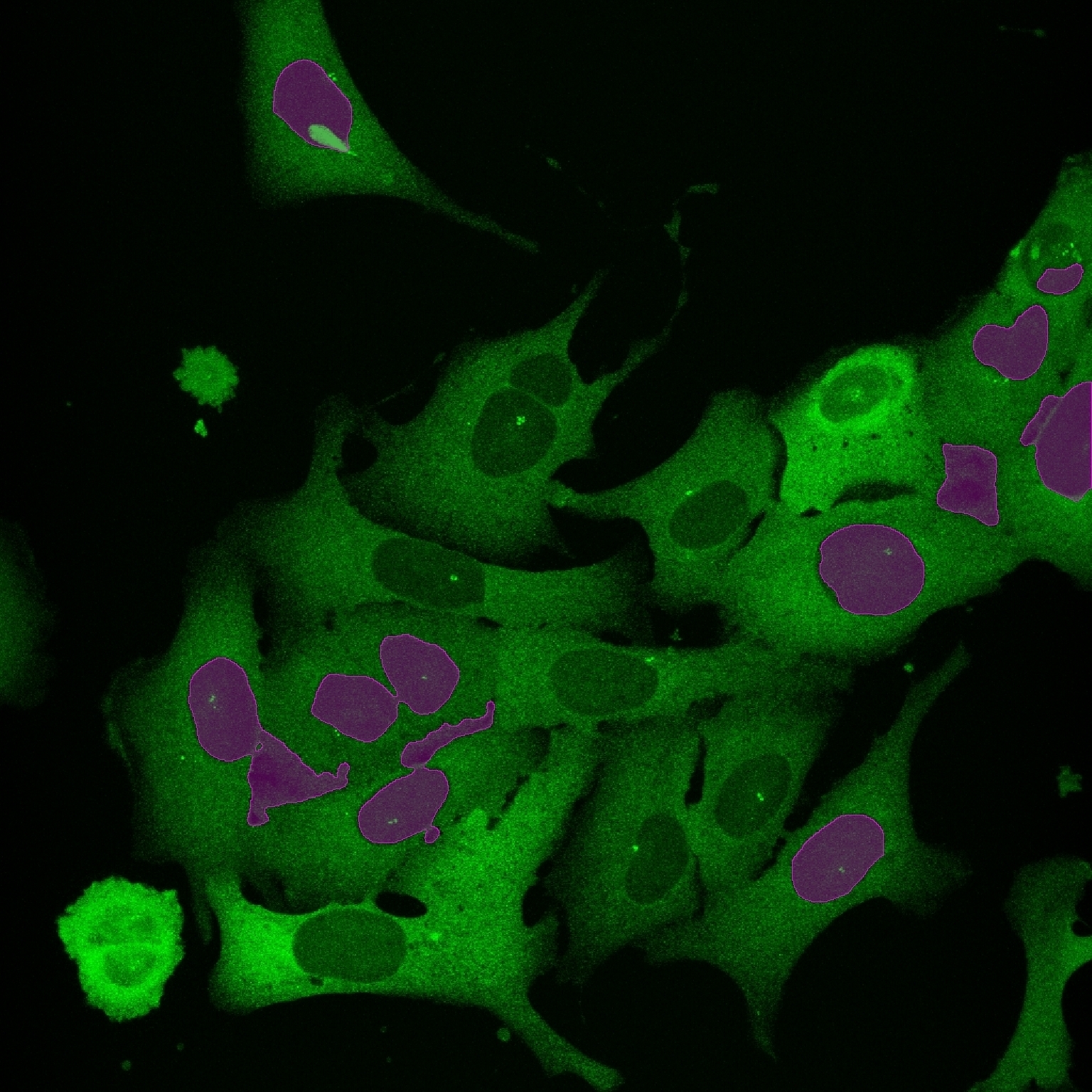 Conventional Thresholding
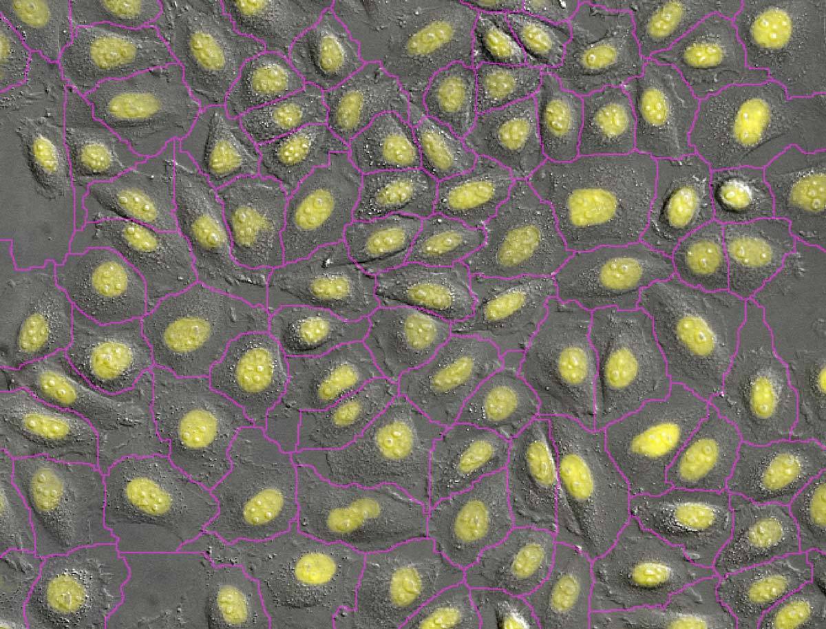
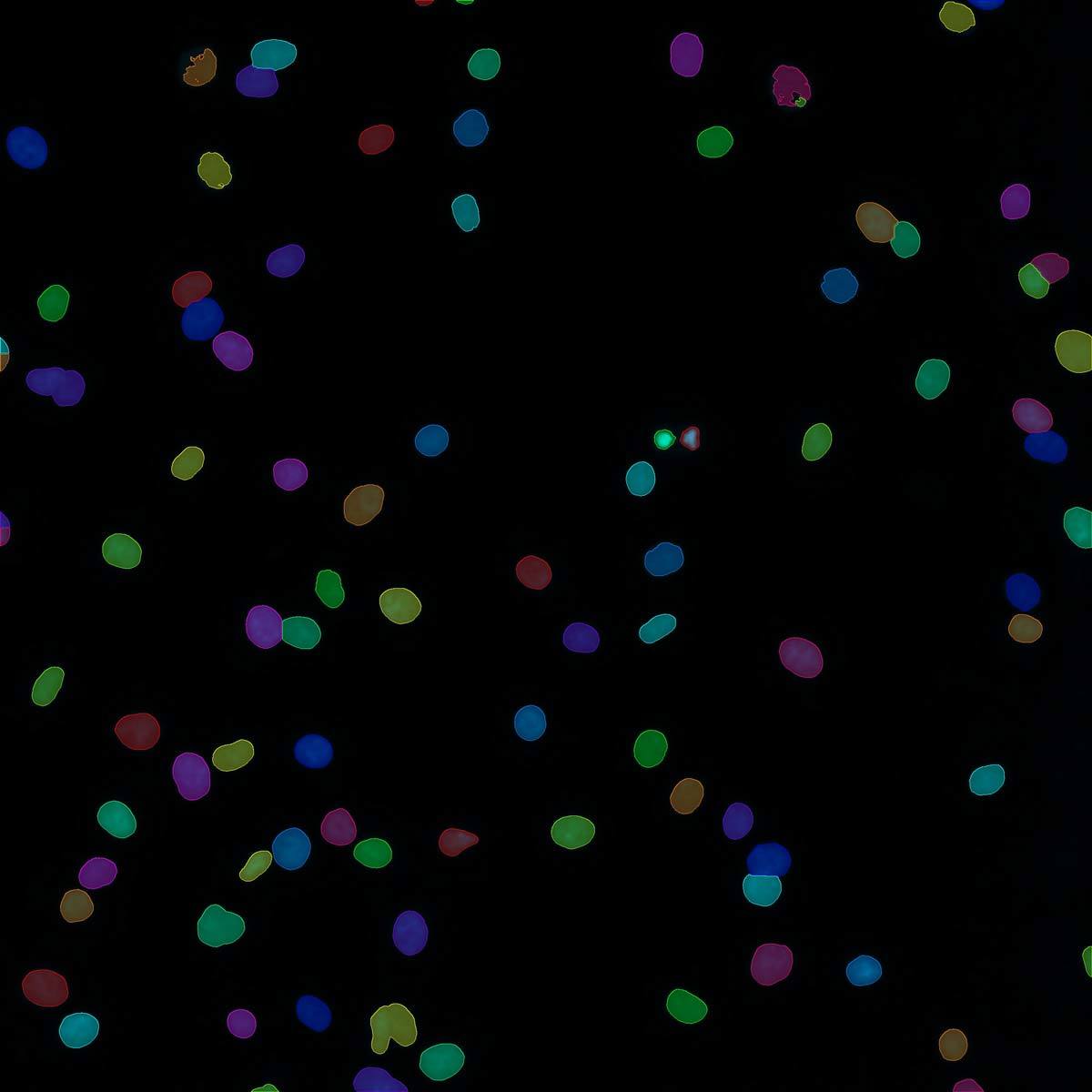
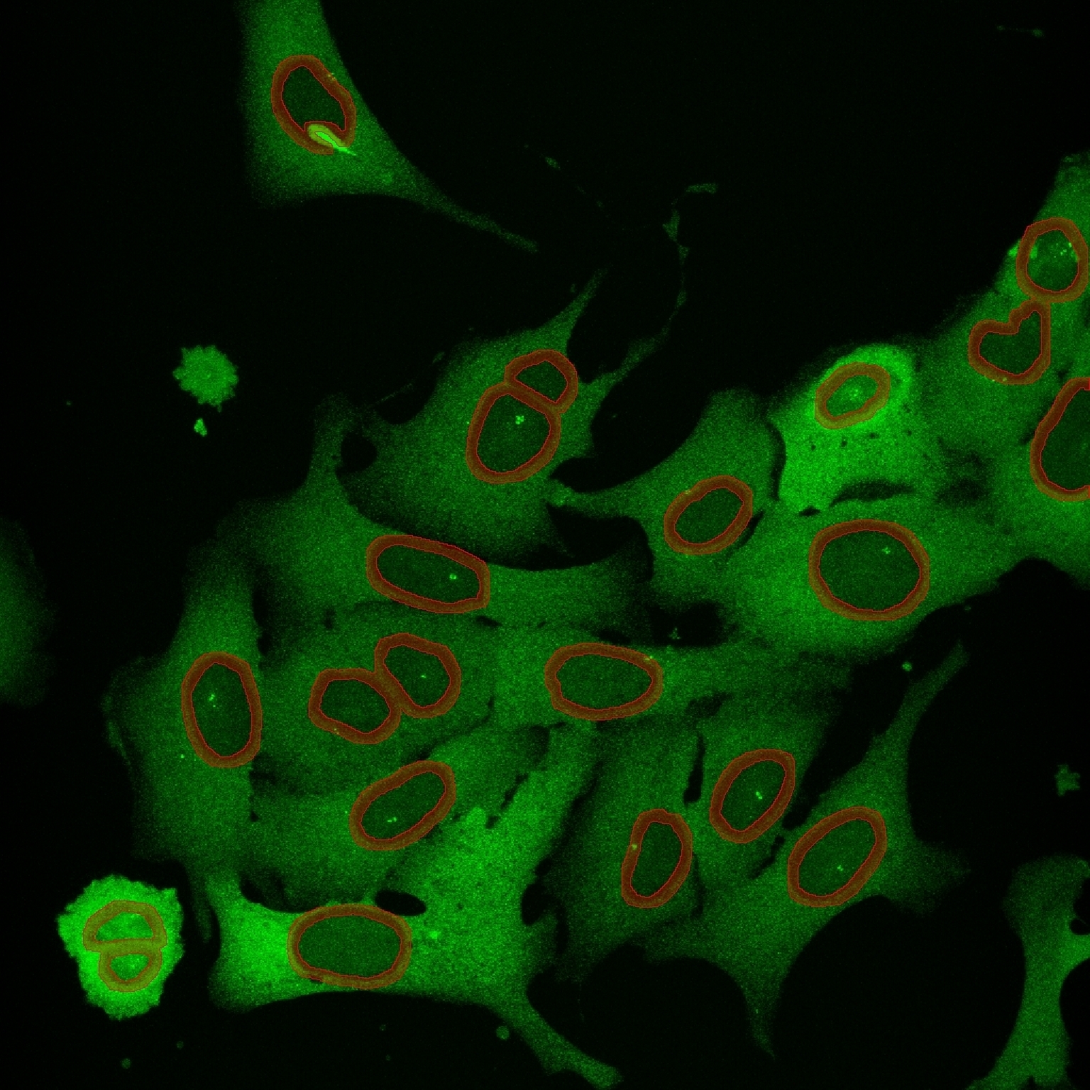 AI Segmentation
Intensity measurements were desired to be made along the nuclear envelope of cells. Conventional segmentation could not differentiate the cellular structures and misses several cells. AI-trained segmentation recognizes and identifies the nuclear envelope successfully.
 Raw Image
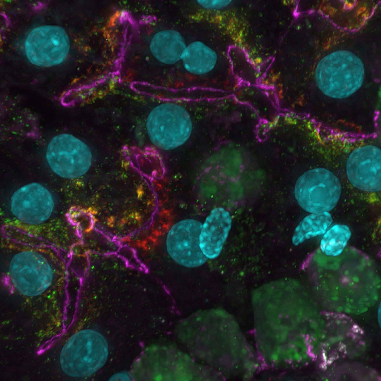 AI
AIWidefield data can be contaminated by scattered and out-of-focus light, but AI-based tools can recover high contrast images by removing noise and blur.
Clarify.ai Module
Clarify.ai uses artificial intelligence to automatically remove blur from fluorescence microscope images.
Clarify.ai utilizes new Nikon technologies executed on graphic processing units (GPUs) to leverage fast and efficient clarity in images normally corrupted by blur due to out-of-focus light.
The module is pre-trained to recognize fluorescence signal emitted from out-of-focus planes and can computationally remove this haze component from the image automatically, leaving behind the in-focus structures, and can be used on any widefield 2D or 3D data set, detector, or magnification, without the need for AI training or introduction of bias from complicated user-settings.
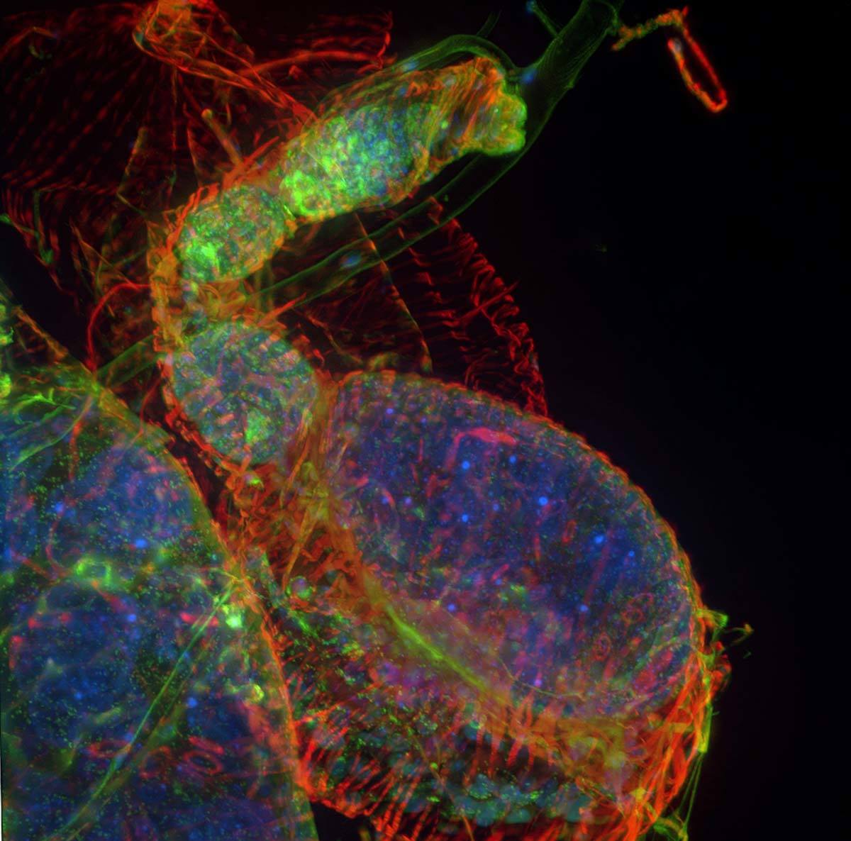 Clarify.ai
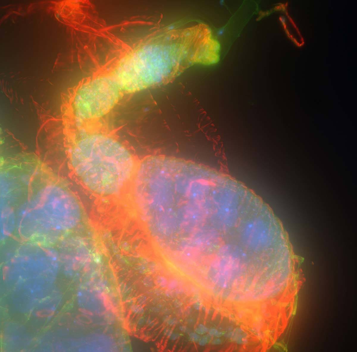 Original
60x Multichannel 3D widefield Z stack before and after application of Clarify.ai
 Clarify.ai
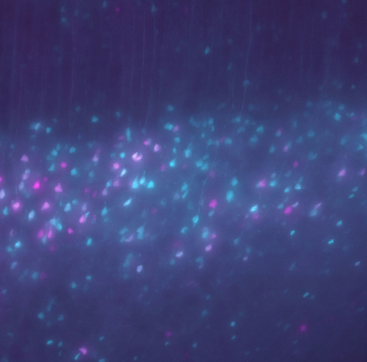 Original
NIS.ai Processing and Analysis Module
The NIS.ai processing and analysis module consists of tools dedicated to improving and enhancing efficiency in data acquisition and simplifying previously complex or difficult analysis routines.
Convert.ai
By recognizing patterns present in two different imaging channels, Convert.ai can be trained to predict what the second channel would look like when only the first channel is acquired.
Commonly, this can be used as a segmentation tool for label-free approaches, or imaging without harmful near-UV excitation. Once the neural network learns the pattern common to two channels, then in subsequent experiments the second channel is no longer needed to be acquired. Throughput of acquisition as well as specimen viability both increase as a result.
 Convert.ai
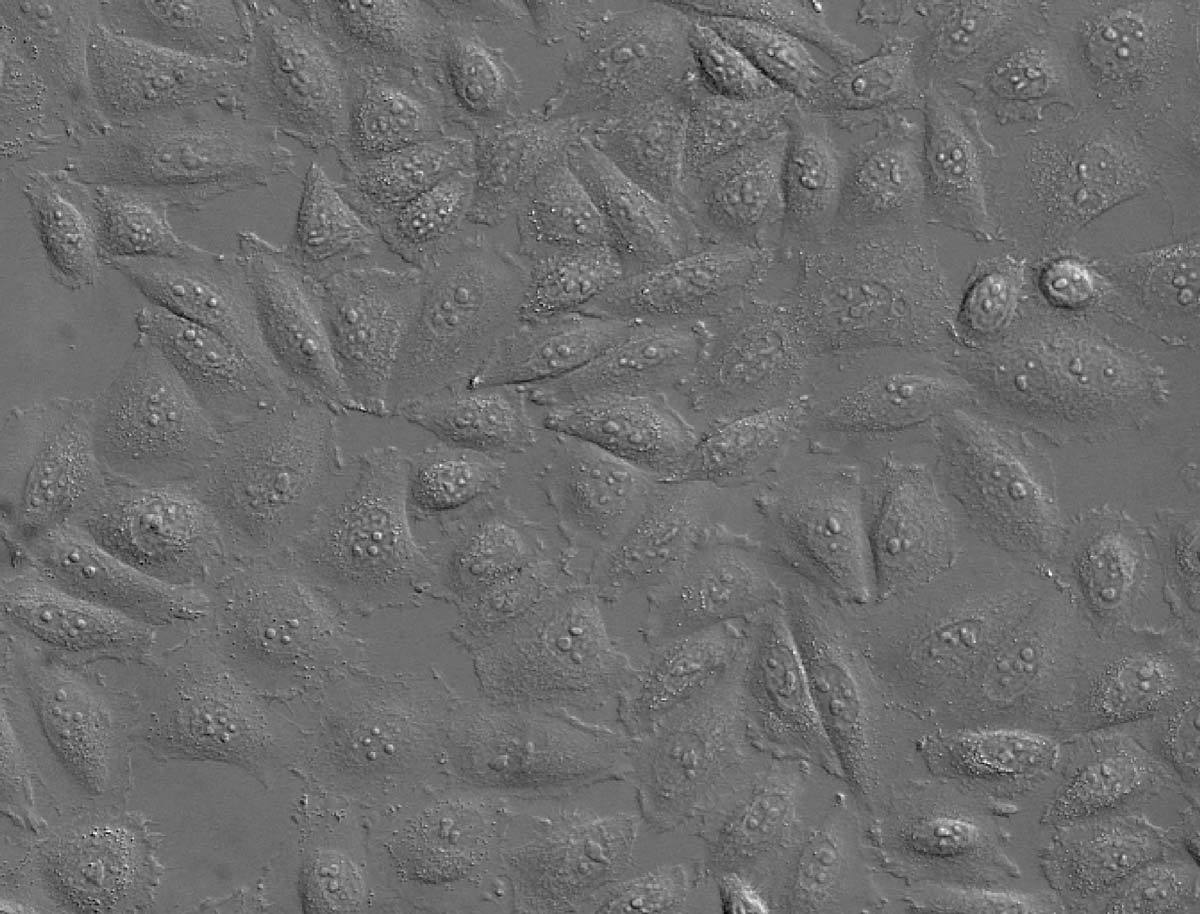 Original
DAPI staining of nuclei is a common method allowing cell counting and segmentation. Convert.ai can be trained to predict where the DAPI label is present in DIC or phase images. This predicted channel can then be used for segmentation and counting, without ever having to label the specimen with DAPI or acquire a fluorescence channel.Photos courtesy of Dr. Kentaro Kobayashi, Division of Technical, Research Institute for Electronic Science, Hokkaido University
Enhance.ai
Some fluorescent samples express a very low signal and it is difficult to visualize or extract details for segmentation.
In addition, many of these samples are sensitive to light or photobleach very quickly and need to be imaged as fast as possible.
Enhance.ai can restore details by training the network what properly-exposed images look like. Then this recipe can be applied to underexposed images to restore detail that can be used for further analysis.
 Enhance.ai
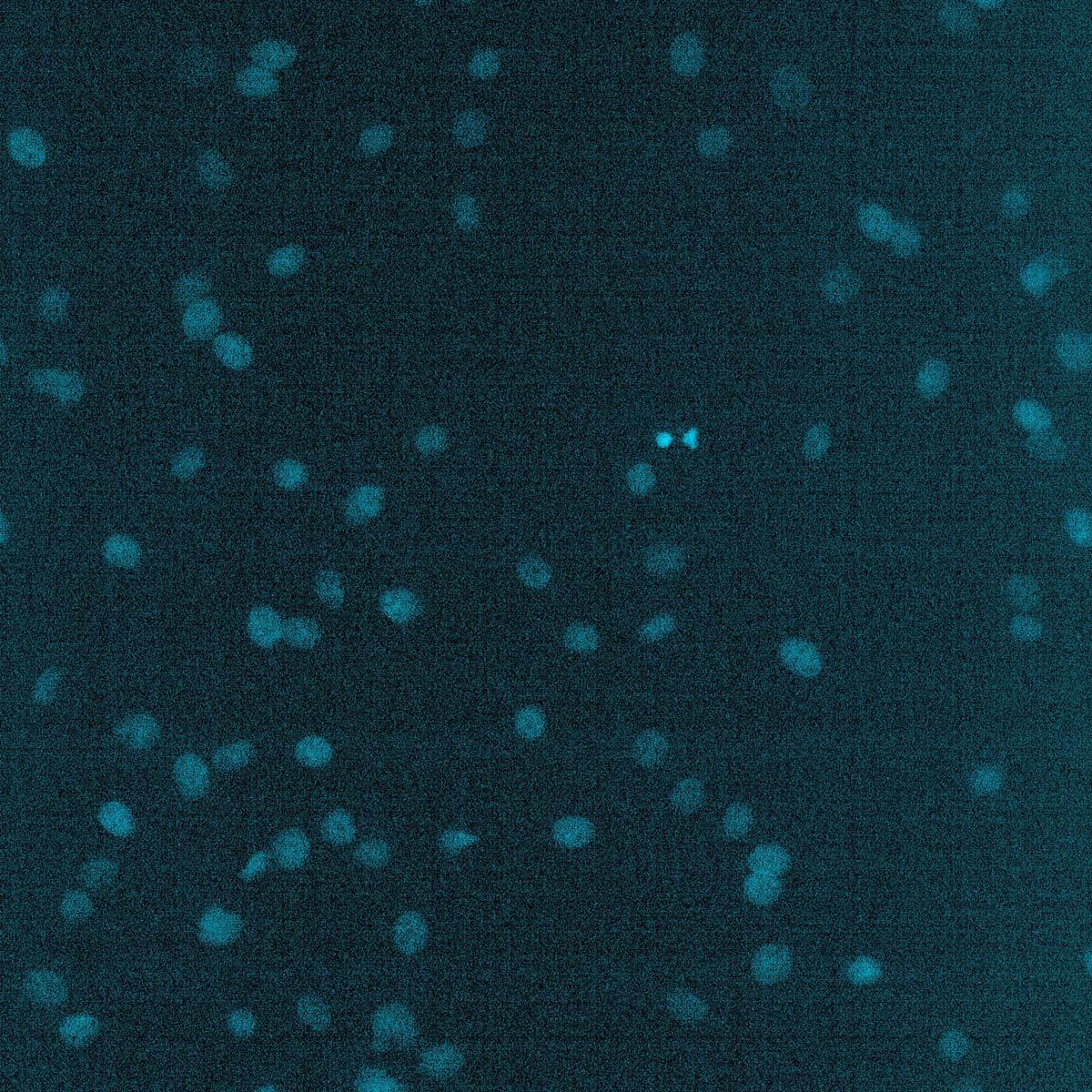 Original
DAPI stained nuclei are purposefully underexposed to limit the specimen’s exposure to near-UV light. Enhance.ai is used to restore the signal-to-noise ratio to normally exposed DAPI staining, for easy segmentation and counting.
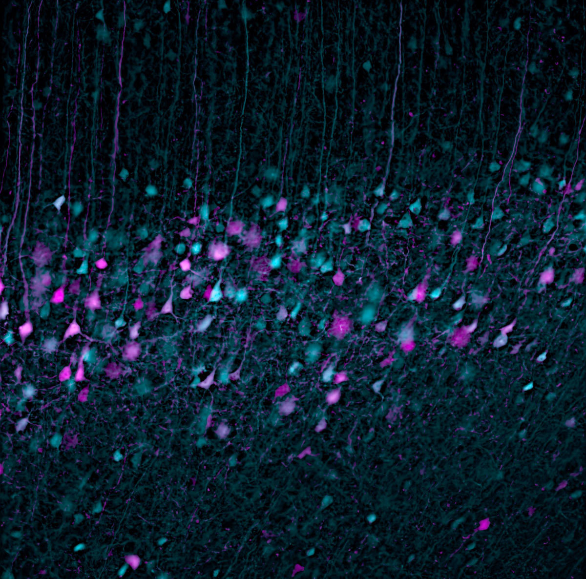
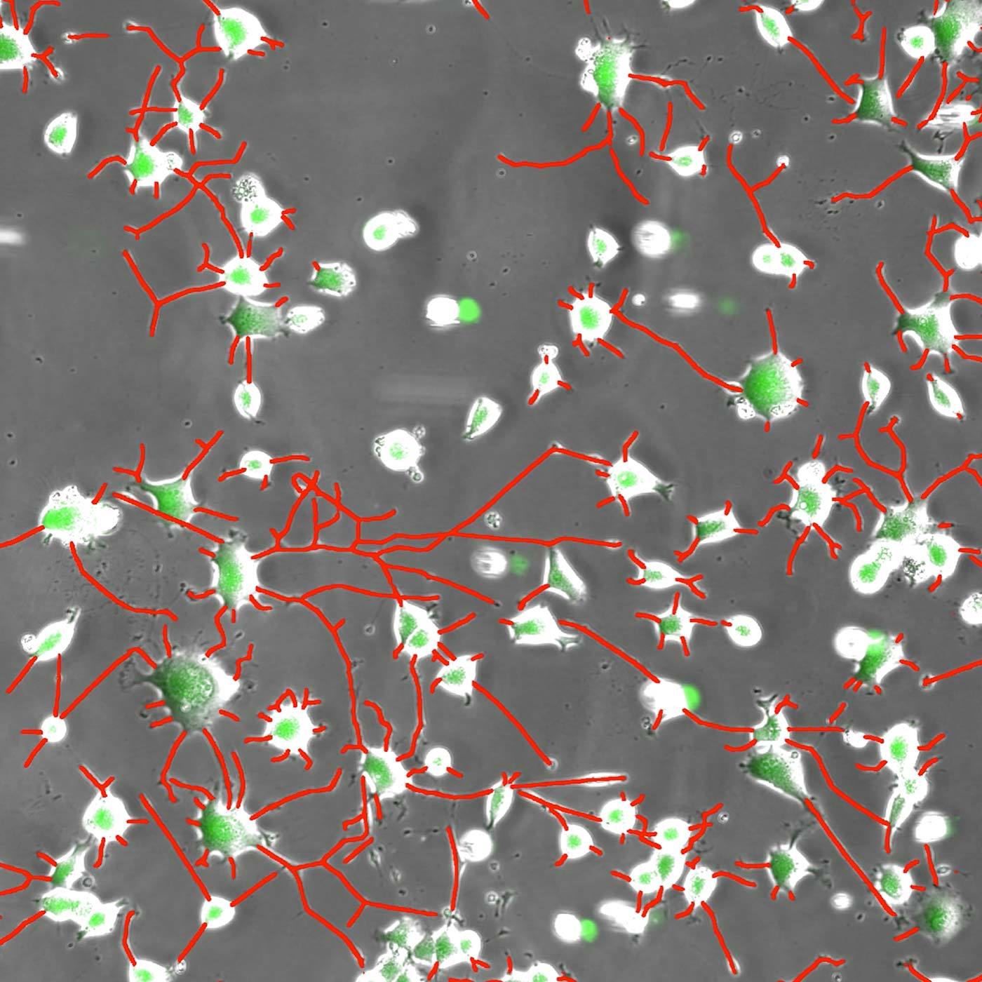
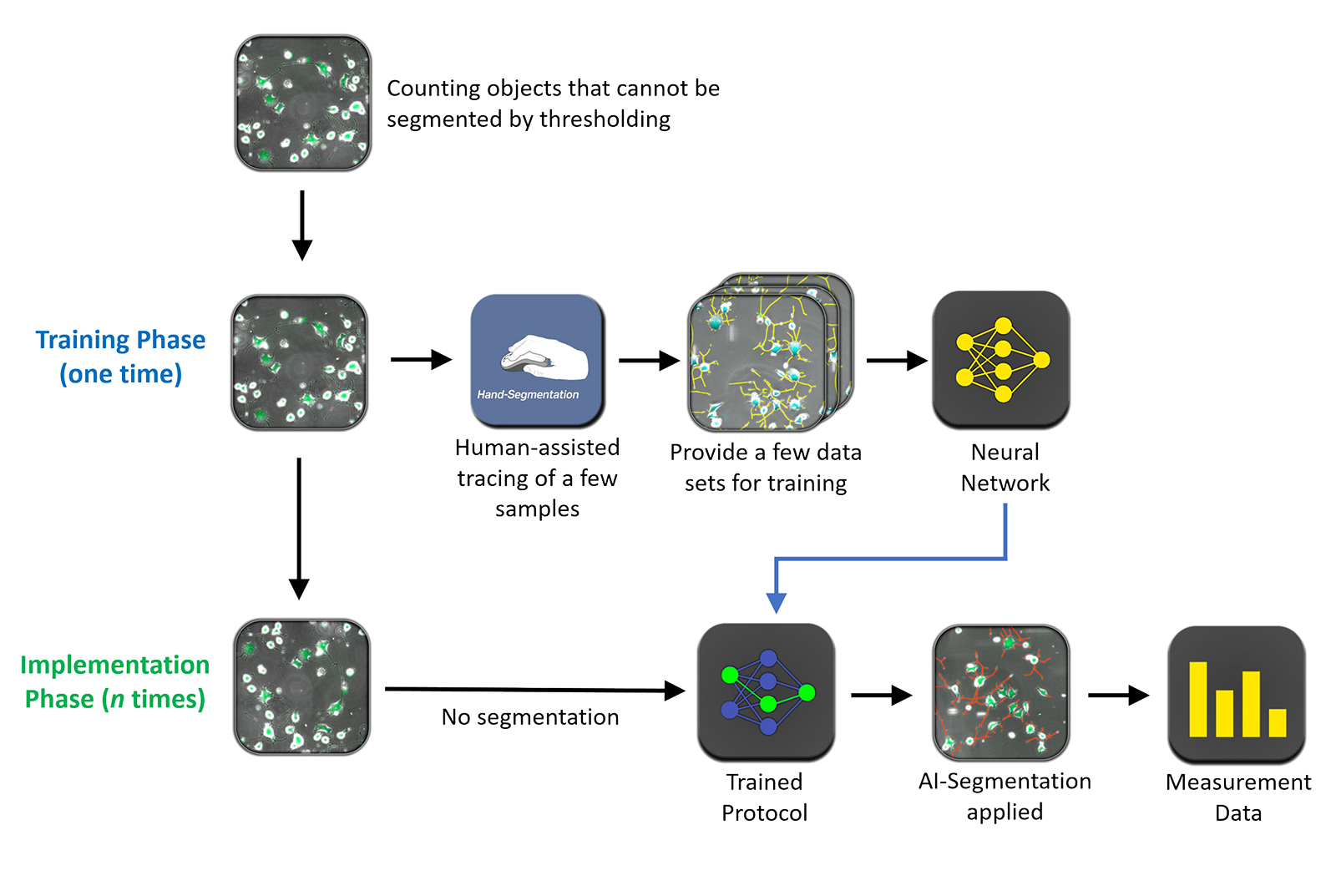
Segment.ai
Some images are nearly impossible to segment by traditional intensity thresholding methods. A neural network can be trained by human classification of structures of interest that cannot easily be defined by classic thresholding and image processing by using Segment.ai.
By tracing features of interest and training these compared to the underlying image, the neural network can learn and apply segmentation to similar images, recognizing features previously only identifiable by painstaking manual tracing approaches.
 Segment.ai
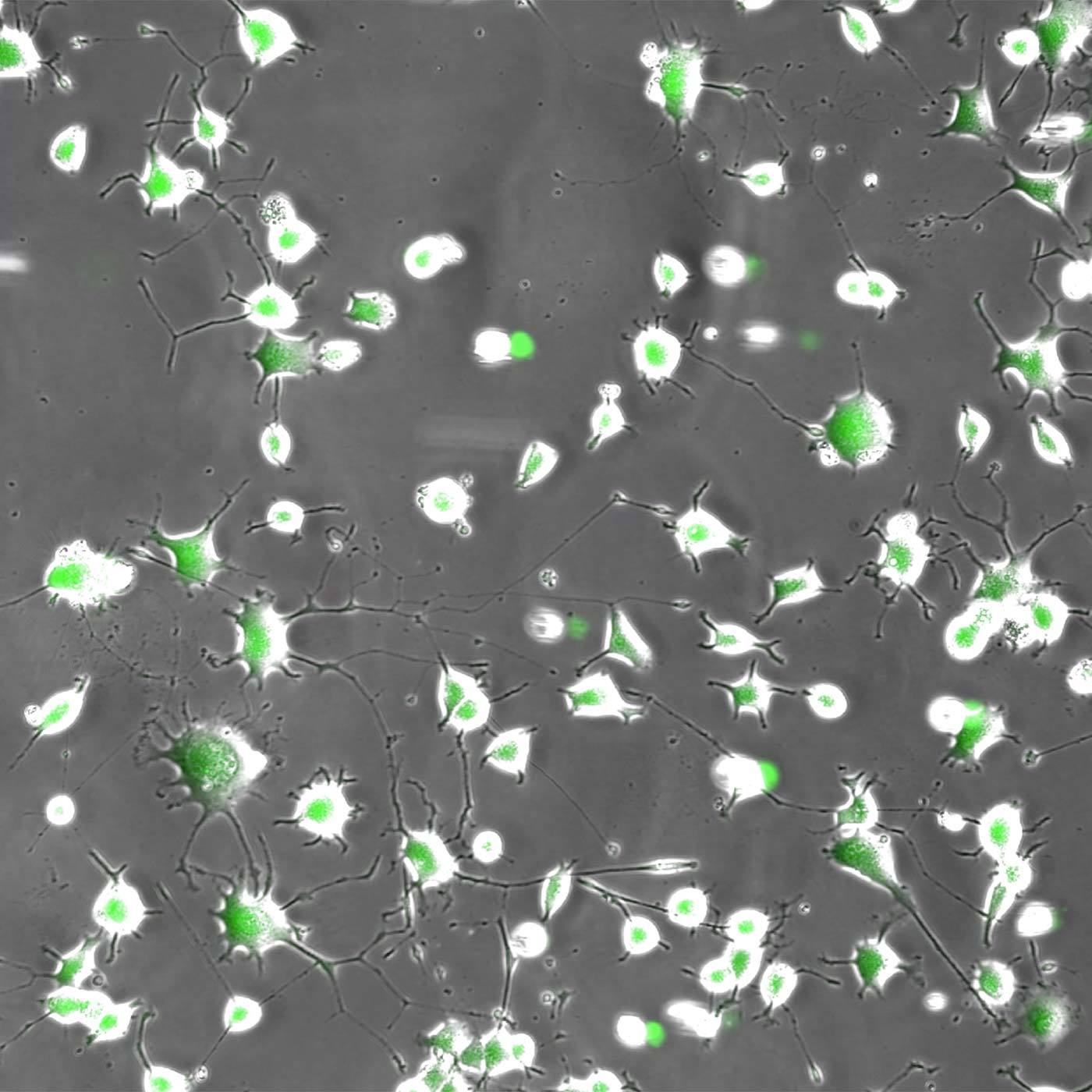 Original
Neurites in phase-contrast were not possible to define accurately by traditional thresholding. Segment.ai was trained on hand-traced neurites (human recognized) and learned how to trace neurites in subsequent images.
Denoise.ai Function
Included in the NIS-Elements AR core package, Denoise.ai can be applied to confocal images to remove shot noise. All images contain shot noise, which is a Poisson-distributed noise related to discreetly sampling (acquiring images) of a continuous event. As signal levels decrease, the contribution of shot noise increases and noisy images result, following a square-root function. Such noise therefore is modeled in a neural network and doesn’t need to be further trained.
With new fluorescent techniques pushing intensities lower and acquisition speeds increasing, Denoise.ai can recognize and remove the shot noise component of images, increasing clarity and allowing for shorter exposure times or more exposures of specimens while maintaining viability.
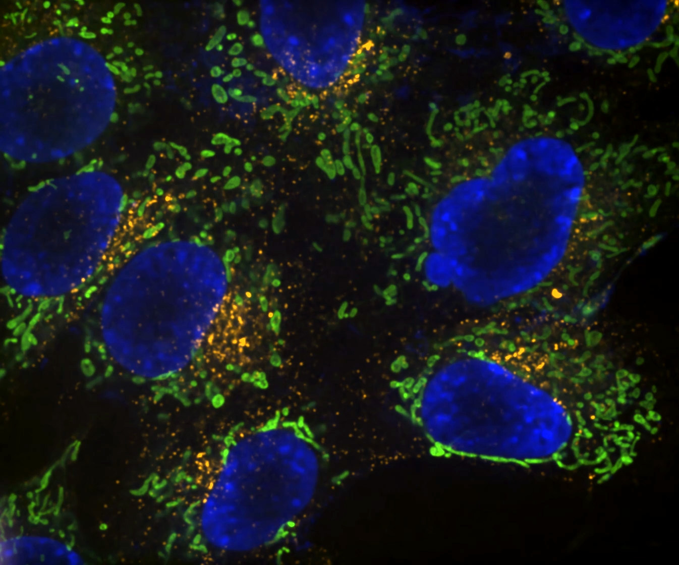 Denoise.ai
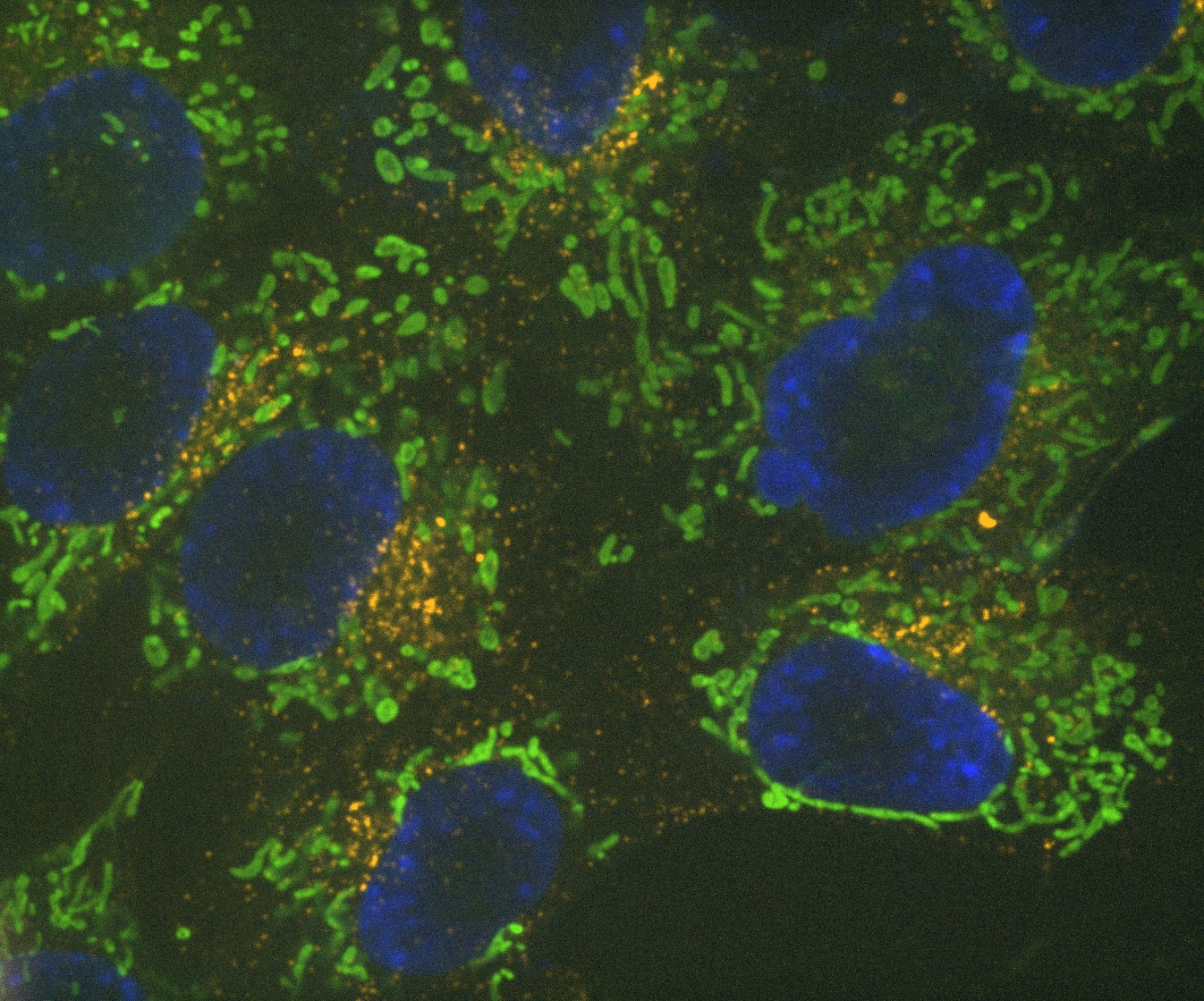 Original
Denoise.ai can be applied to remove the shot noise component of images while leaving the underlying structure and intensity values undisturbed.
No programming skills required
Clarify.ai and Denoise.ai are pre-trained deep learning networks and require no additional settings or parameters in order to apply these tools automatically to images.
The NIS.ai Processing and Analysis module uses training data to specifically target and address user-defined experiment parameters: it employs convolutional neural networks (CNNs) to learn from labeled training data created by either conventional segmentation or human-assisted tracing of small subset of representative samples.
When using the module, the software interface makes it easy to apply complex deep learning to sample data, eliminating the need to design a complex neural network and apply training data to it.
Automated tools take this training data and apply the neural network to recognize patterns. The result training recipe can then be applied repeatedly and reliably to similar samples to process or analyze huge volumes of data at significantly faster speed than traditional techniques.
GA3: an analysis pipeline with AI capabilities
Using NIS-Elements General Analysis (GA3), multiple conventional segmentation and AI tools can be combined to create data measurement routines customized for a specific experiment. These can be applied across multiple images, experiment runs, or high content data.
Because GA3 is freely customizable, it can be adapted to new experiment routines easily. Routines can be embedded as well during experiment acquisition runs.
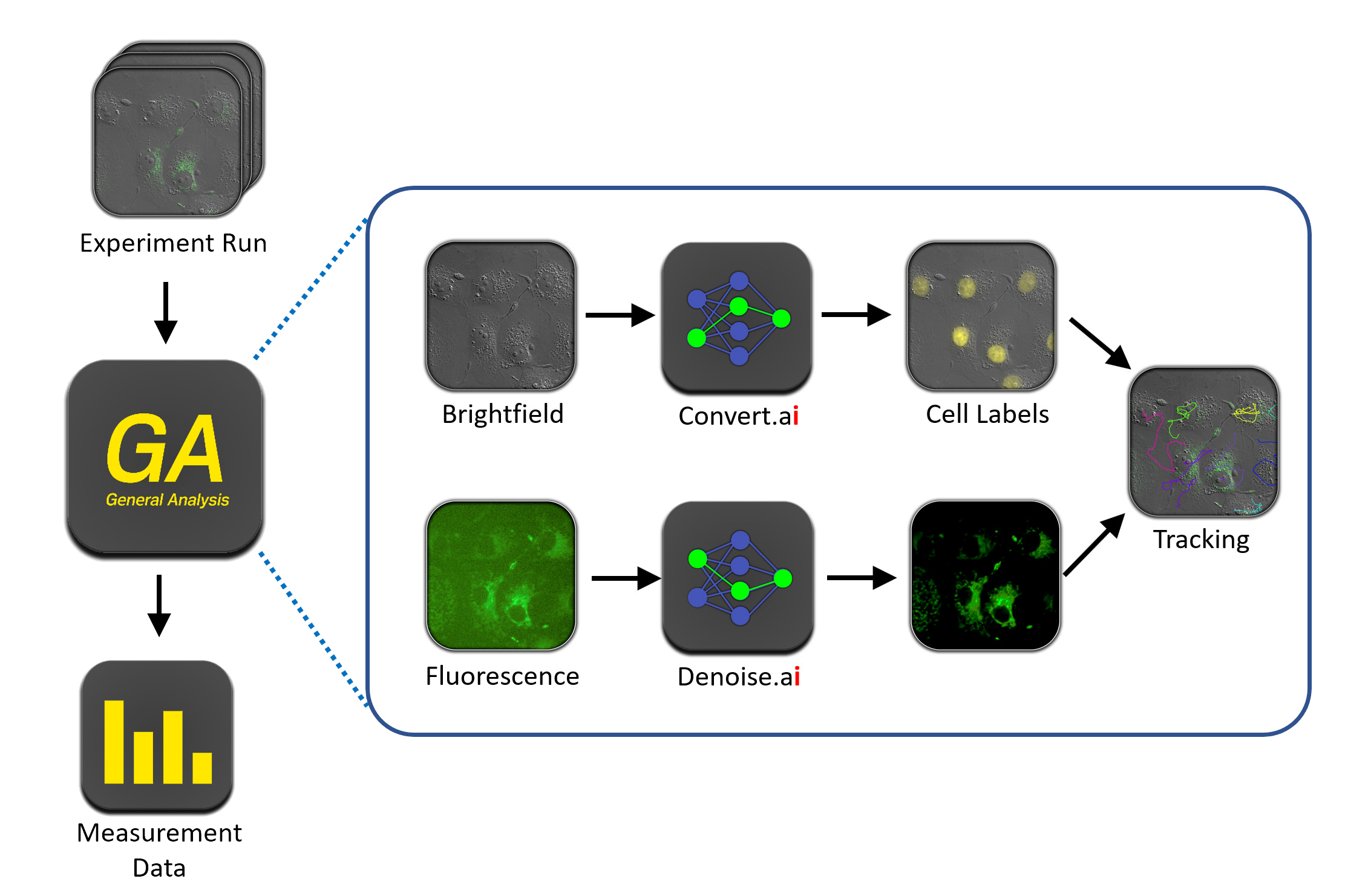
General Analysis is used to apply Convert.ai to brightfield images to mark nuclei, and Denoise.ai applied to a noisy fluorescence channels. The converted channels can then be tracked over time lapse to measure cell movements. This routine is then applied to multiple data sets for data measurement.
Use NIS.ai as part of an imaging pipeline
NIS.ai tools can be combined with all other features of the NIS-Elements platform to develop imaging protocols and targeted analysis from basic counting through rare event or selective phenotype detection and analysis.
This can be incorporated post-acquisition, or more impactfully, as an integral part of an experimental protocol so that NIS-Elements Intelligent Acquisition analysis results obtained during the experiment run guide the experimental parameters in different directions.
Using the JOBS experiment wizard, customized experiments with embedded analysis tasks and branches based on analysis results can be created, allowing for higher throughput and more targeted acquisitions.
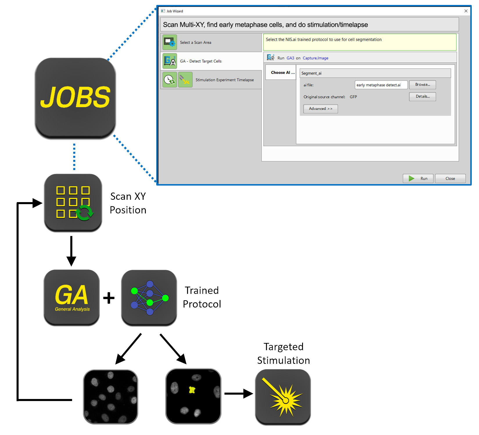
Example of utilizing Segment.ai in an experiment run to analyze XY positions as they are captured, and to search for specific phenotypes. When a target cell is found, a stimulation experiment is performed. If no target cell is found, the experiment proceeds to the next XY position.
Quantifiable Results
Artificial intelligence has become commonly accepted in diagnostic imaging and is an increasingly popular tool for a number of applications. Its appeal over traditional mathematical approaches is both its speed and incredible accuracy. However, it is important to be able to validate the results of AI computations, and to utilize these results appropriately for computational analysis.
NIS-Elements software provides feedback during training routines to indicate the confidence of the trained neural network to provide accurate results, as well as several analysis tools and workflows to validate the efficiency of the neural networks, or to allow easy comparison of AI data to ground truth data.
Summary of AI Modules
●: included, ⚬: option
| Denoise.ai | Clarify.ai | Enhance.ai | Segment.ai | Convert.ai | |
|---|---|---|---|---|---|
| NIS C, Ar, Ar ML, Ar Passive | ● | ⚬ | ⚬ | ⚬ | ⚬ |
| NIS.ai Module | ● | ● | ● | ||
| Deconvolution Modules | ● | ||||
| Batch Deconvolution | ● | ||||
| Offline Batch Denoise Package | ● |
NIS-Elements Advanced Research
Nikon’s Flagship NIS-Elements Package
Optimized for advanced research applications, Nikon’s flagship software package features fully automated image acquisition, advanced device control and powerful analysis and visualization tools.
Acquire
- From Single to Multi-Dimensional imaging
- Intuitive display of multi-dimensional datasets with multiple viewing options (e.g. slice, volume, time, spectral, binary, etc.), easy data management, and export-ready
- Go Beyond Image Capture
- Photo-Stimulation
- Perfusion
- Realtime measurements (e.g. Ca2+, FRET) during image acquisition
- Image without Constraints or Limitations with JOBS
- Custom-build your acquisition workflow with graphical programming
- Intelligent Acquisition with JOBS and General Analysis (GA)
- Let the results dictate the acquisition parameters with conditional workflows
- Only acquire meaningful data
- Reduce time spent on data mining
- Maximized Device Performance
- NIS-Elements is designed to optimize the speed at which all components can perform
- Maximize device usage through hardware triggering
- Enables fastest possible image capture times for imaging and tracking fast biological events
- Eliminates unnecessary light exposure, extending the health of the sample for long-term time lapses
- Largest 3rd party integrator in the industry
- Easily integrate and control non-Nikon devices for fully customized experiments
- Quickly adapt to changing research needs by utilizing 3rd party devices
Quantitate
- From simple manual measurements to automatic data collection of hundreds of parameters
- Full suite of binary and image processing tools in General Analysis (GA)
- Multi-channel binary software assay toolbox
- 2D and 3D Tracking and Measurement
- Realtime analysis of live cell dynamics during acquisition
- Intensity Based Measurements
- Morphological Based Measurements
- Object Tracking
Process and Visualize
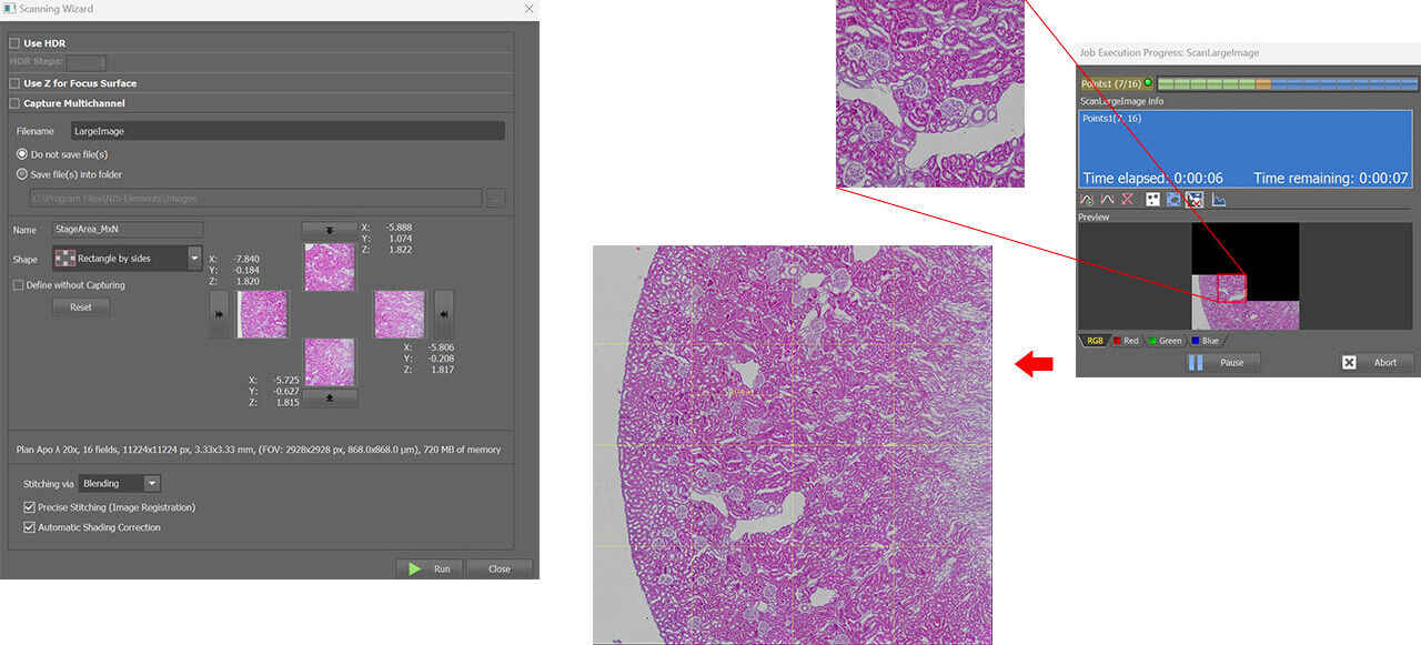
- Easily view tiled/stitched large images
- Unique Extended Depth of Focus (EDF) view for creating beautiful, high-contrast, in-focus 2D projection images from 3D data (option)
- Advanced 3D and 2D deconvolution algorithms for extending resolution
- Unique visualization options such as XYT view (3D kymograph)
Publish
- Mulitple Image formats
- Advanced movie creator with annotations
- Easily combine images and analysis results to create beautiful figures
- Data export to Excel, Matlab or any third party or customer software
NIS-Elements Basic Research
Microscope imaging software geared for acquisition and device control for standard research applications, requiring four-dimensional imaging.
Developed for standard research applications such as analysis and photodocumentation of fluorescent imaging, NIS-Elements BR features up to four-dimensional acquisition and advanced device control capabilities.
- Basic Multidimensional Imaging
- Integration of all moving parts so you can focus on science and data
- Measurement
- Intensity, ROIs, Counting, Morphology
- Large Image Stitching
- Show Multi-Channel Data & Timelapse
- Unique Extended Depth of Focus (EDF) view for creating beautiful, high-contrast, in-focus 2D projection images from 3D data (option)
NIS-Elements D
Nikon’s Photodocumentation NIS-Elements Package
Software package for photo-documentation. Includes basic measuring and reporting tools.
* NIS-Elements D is not for clinical diagnostic use.
Easy capture, processing, and storage
- Easy camera controls and presets
- Flexible imaging: Automated XY, Z control, or Timelapse
- Manual and Automated Measurement
- Counting and morphology
- Annotate your datasets
- Unique Extended Depth of Focus (EDF) view for creating beautiful, high-contrast, in-focus 2D projection images from 3D data (option)
- High Dynamic Range (HDR) image acquisition combines images acquired with different exposure times to capture the full dynamic range of the specimen in one image (option)
- Supports custom workflow using macros
- Single document interface. Does not support fluorescence imaging nor wavelength switching
NIS-Elements Confocal & Enhanced Resolution
Nikon’s Confocal NIS-Elements Package
Dedicated interface for Nikon’s confocal and multiphoton systems, providing easy instrument setup and streamlined operation. Incorporates many of the features of NIS-Elements AR for advanced acquisition, image processing, analysis, visualization and data sharing capability.
Resolution
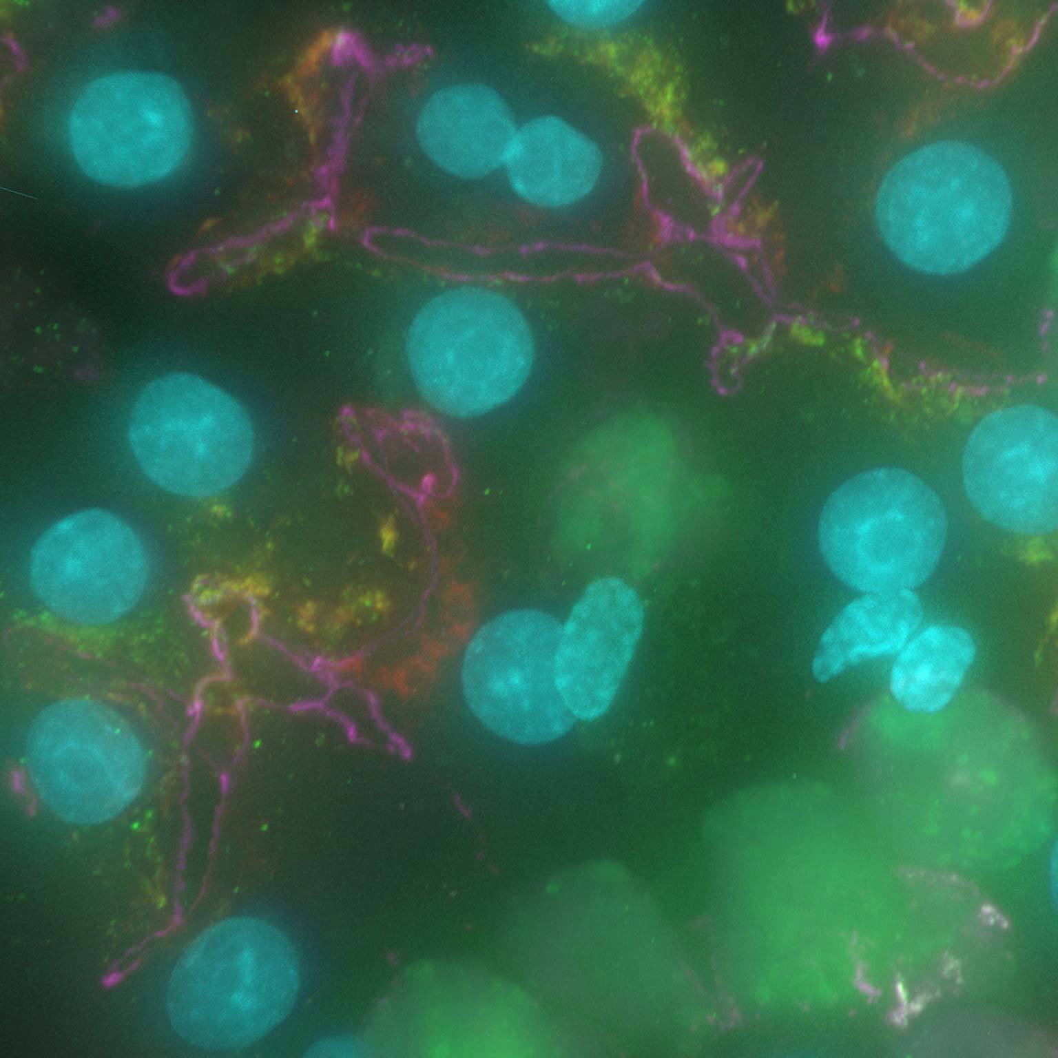
Image resolution is defined as the smallest distance between 2 points that can be resolved. The theoretical limit of resolution for a conventional optical microscope is approx. 200 nm. Higher resolution images can be theoretically achieved with confocal microscopes, but this has not been effectively achieved. Resolution can be increased beyond the conventional confocal image resolution (up to ~1.5 times improvement in XY; up to ~1.7 times improvement in Z) by using specific image acquisition and processing techniques.
 Standard confocal image
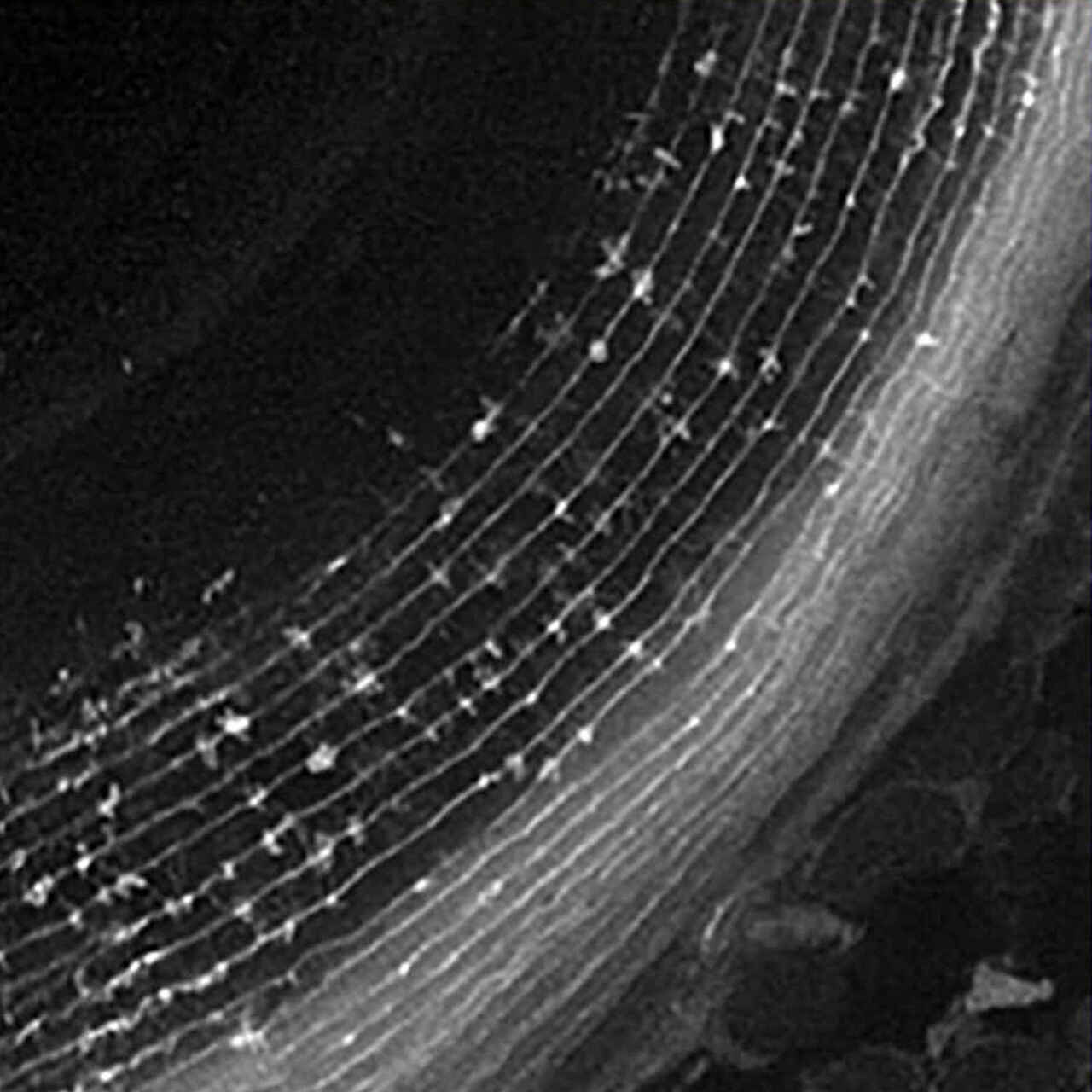 Enhanced Resolution image
Image courtesy of: Drs. Toshiaki Mochizuki and Ichiro Masai, Developmental Neurobiology Unit, Okinawa Institute of Science and Technology Graduate University

Standard confocal image

Enhanced Resolution image
Red: Central spindle, Blue: Nuclei
Image courtesy of: Toshinori Hyodo Ph.D., Department of Biochemistry, Aichi Medical University School of Medicine
 Standard confocal image
 Enhanced Resolution image
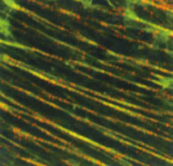
Stress Fibers (LLC-PK1, Pig Kidney Cells), Green: F-actin, Red: Myosin Heavy Chain
Image courtesy of: Keiju Kamijo Ph.D., Division of Anatomy and Cell Biology, Faculty of Medicine, Tohoku Medical and Pharmaceutical University

Standard confocal image

Enhanced Resolution image
Apical surfaces of auditory epithelia of mouse cochleae were stained by Atto-565-phalloidin at postnatal day 2.
Image courtesy of: Dr. Hideru Togashi, Division of Molecular and Cellular Biology, Department of Biochemistry and Molecular Biology, Kobe University Graduate School of Medicine
NIS-Elements HC
High Content Analysis option
Total acquisition-to-analysis solution for high-content imaging applications. Seamless workflow from microscope and peripheral device control to data analysis and management.

Speed and Flexibility
Streamlines high-speed, automated well-plate acquisition, data review, analysis and management of multiple well plate experiments.
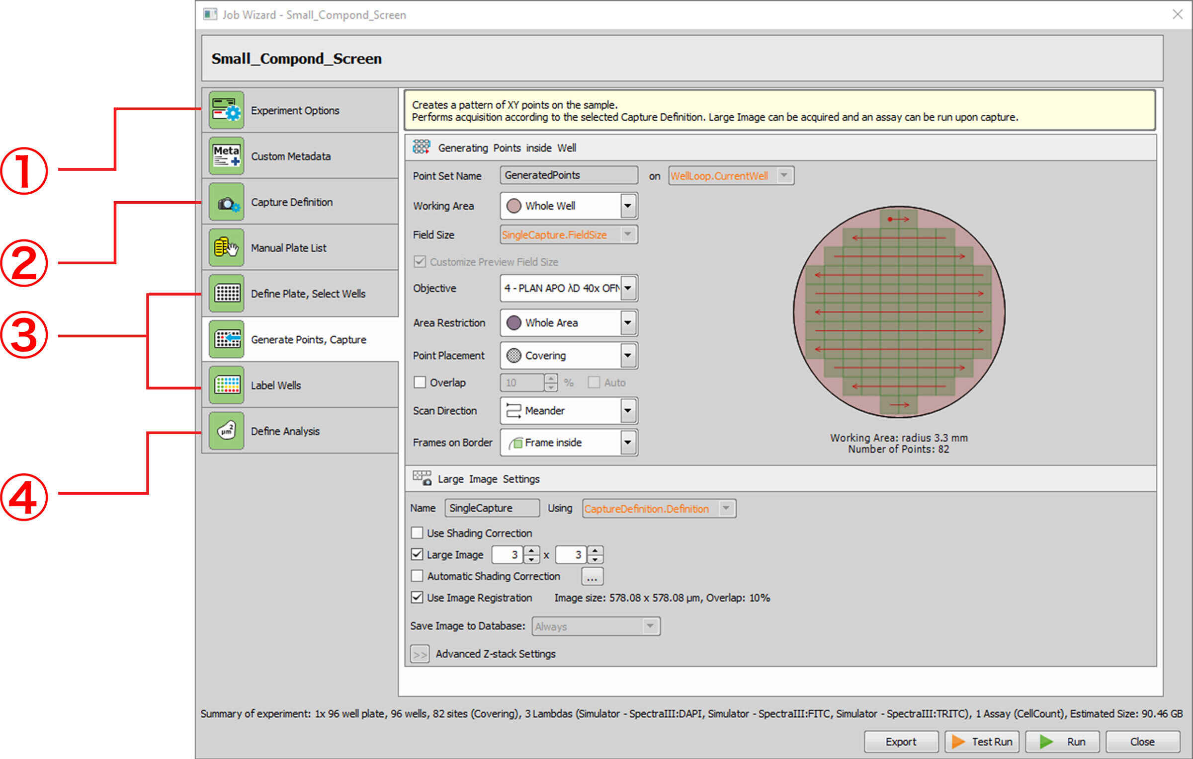
NIS-Elements HC interface simplifies experiment setup using wizards. Easily define acquisition parameters including well-plate configuration, plate handling, autofocusing, filter switching and detectors.
① Define general job parameters
• Z-stack
• Sample labelling
• Autofocus
• Sending task completion by e-mail or SNS
② Define optical configurations for image capture
③ Well plate setting
• Define well plate to use
• Select well plate for image capture
• Define XY image capture pattern inside a well
• Sample labelling
④ Define analysis
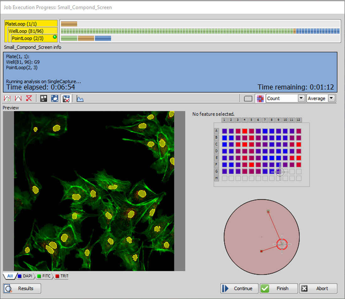
Review
Realtime viewing of data acquisition and analysis progress for instant inspection. Multiple analysis assays can be run simultaneously during the imaging phase or run post-acquisition on offline stations.
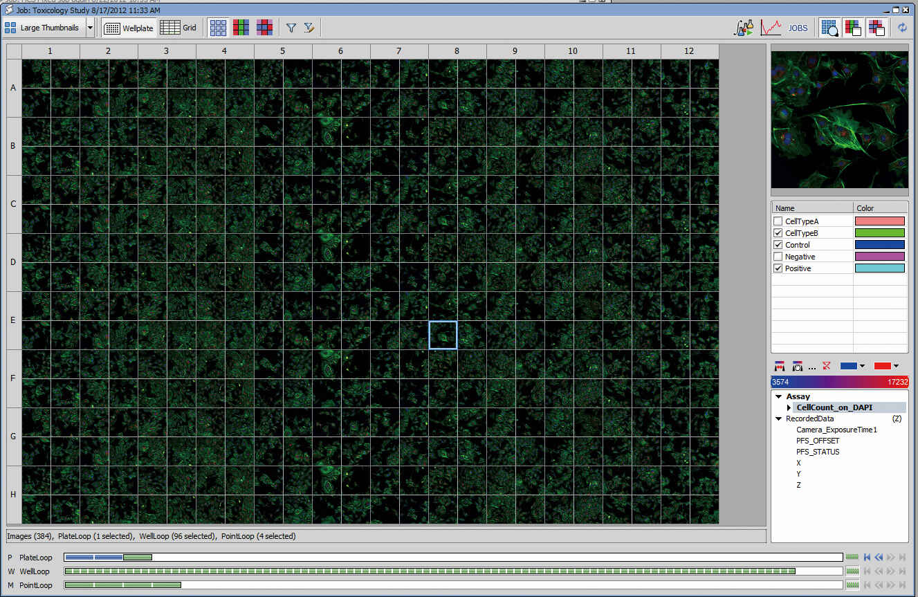
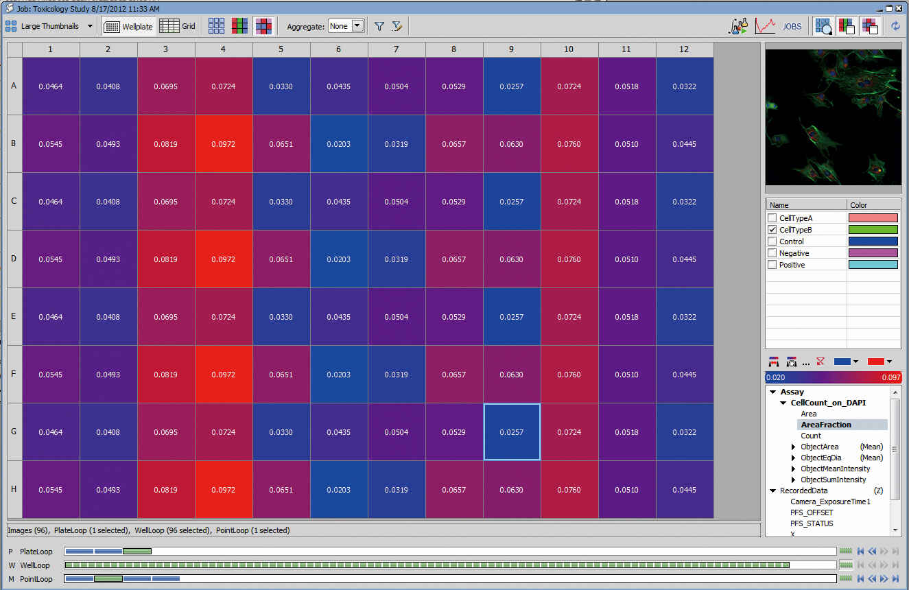
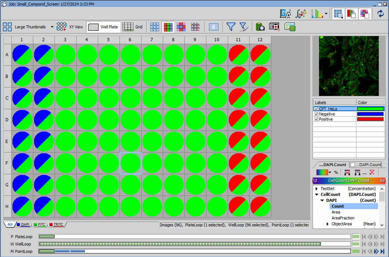
Analysis
Heat Maps of well plates, sample images, binary masks, assay results, sample labels and other metadata are centralized for quick filtering, gating and drill down to cellular detail.
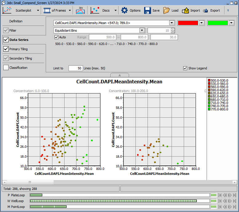
Graphing
Create graphs instantly for data review. Classify, filter, tile, label data points from several different graph types. NIS-Elements offers histogram, scatterplot, bar chart, XY line, classification and gating functions. Easily navigate within the Plate View and export to Excel or bitmap.