
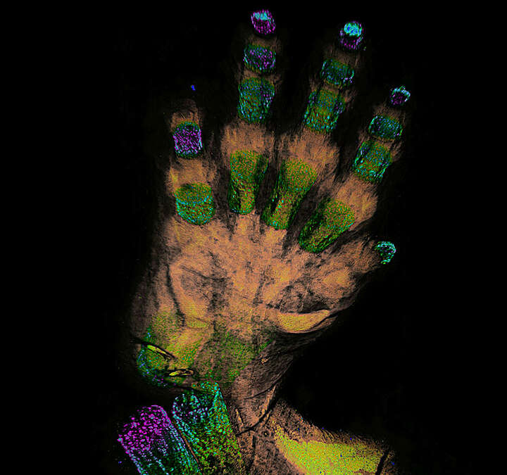
Overview & Features
Nikon’s multiphoton confocal microscopes, which clearly visualize fine structures deep within living organisms, have evolved even further. The AX R MP is equipped with a high-speed resonant scanner with 2K resolution and can capture in a single scan dynamics that span a wide area with superior spatial and temporal resolution.
In addition, the innovative NSPARC super-resolution detector utilizes a newly developed SPPC array detector to collect a two-dimensional image at each scanned point, achieving a significant improvement in resolution. This enables macro-to-micro imaging with a single microscope system.
The AX R MP maximizes image size, as well as spatial and temporal resolution, providing a uniquely powerful solution for any multiphoton experiment.
Features
Nikon’s multiphoton confocal microscopes, which clearly visualize fine structures deep within living organisms, have evolved even further. The AX R MP is equipped with a high-speed resonant scanner with 2K resolution and can capture in a single scan dynamics that span a wide area with superior spatial and temporal resolution.
In addition, the innovative NSPARC super-resolution detector utilizes a newly developed SPPC array detector to collect a two-dimensional image at each scanned point, achieving a significant improvement in resolution. This enables macro-to-micro imaging with a single microscope system.
The AX R MP maximizes image size, as well as spatial and temporal resolution, providing a uniquely powerful solution for any multiphoton experiment.
Bigger pictures. Fast.
Featuring a 22mm field-of-view for both resonant and galvano scanners, the AX R MP captures more data in every frame at any magnification. This means less time spent imaging large specimens and more information in every frame.
See more at high magnification
Use the AX R MP field of view and Nikon’s world-class objective lenses to capture large specimens in one image. With resolutions up to 8K x 8K, these images can be captured without sacrificing a single detail.
Redesigned optics maximize performance over a large area and deliver seamless image stitching at much higher speeds. These improvements save time during acquisition and minimize post-processing.
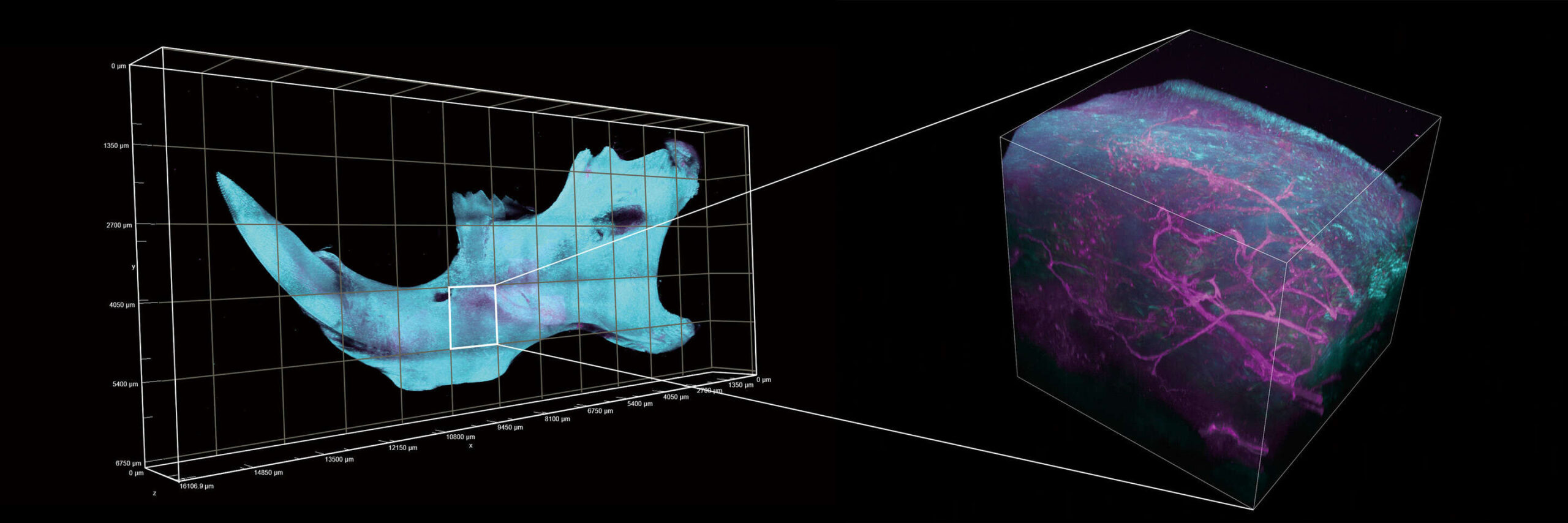
Z-stack image acquired from a 12 x 5 stitched image in 6.7 x 16.2 mm area. Image courtesy of Lin Daniel, PhD, SunJin Lab Co.
Objective: CFI Plan Apochromat Lambda D 10
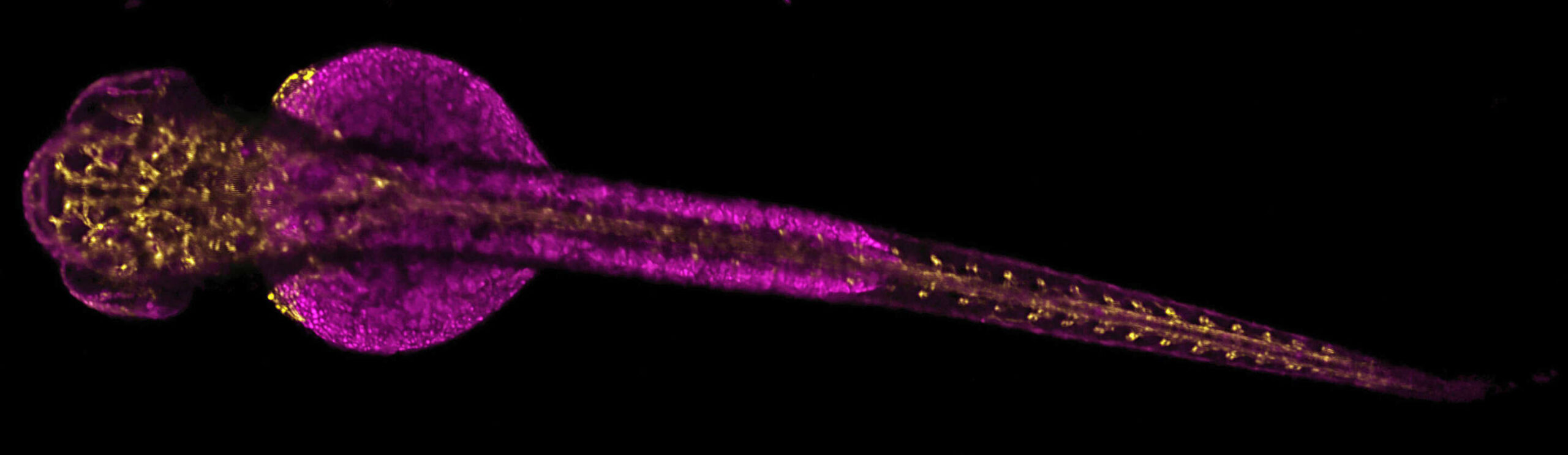
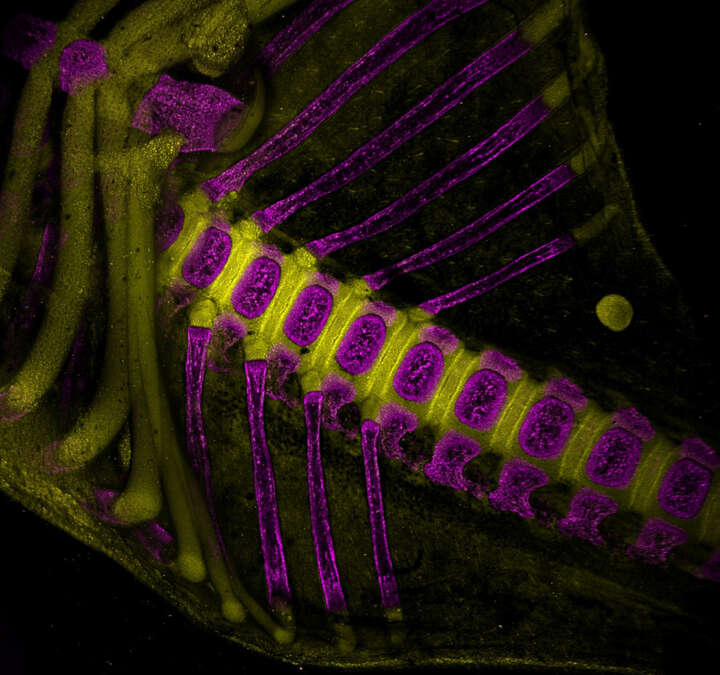
Video of the 3D distribution of blood vessels in a 700 µm thick Z stack acquired in an embryonic zebrafish labeled with GFP and RFP.
Acquired with a CFI75 Apochromat LWD 20XC W objective lens.
Courtesy Erika Dreikorn and Dr. Beth Roman, Department of Human Genetics, University of Pittsburgh Graduate School of Public Health
High-speed imaging that captures the dynamics of life
The 2K x 2K resonant scanner featured on the AX R MP provides fast (up to 720 fps), gentle and high resolution imaging. This means samples live longer and stay healthier all while much higher-quality data is being collected. Alternatively, the resonant scanner can be applied to large fixed samples to dramatically improve the speed of large volume acquistions, with no loss of image integrity.
Individual blood cells are identified in high resolution, and blood flow is imaged at a high speed of 28 fps (2048 x 546 pixels)
Images courtesy of Erika Dreikorn and Dr. Beth Roman, Department of Human Genetics, University of Pittsburgh Graduate School of Public Health
Objective: CFI75 Apochromat LWD 20XC W
Bright, high-definition imaging of deep structures
Utilizing high-resolution resonant scanning, extremely low-noise detectors and efficient multiphoton excitation, the AX R MP collects fluorescence from deep within any specimen for bright, high-definition results.
High resolution deep imaging for intravital microscopy
The AX R MP’s two selectable scanners, resonant and Galvano, allow users flexibility in acquisition, and provide both high-speed and high-resolution solutions. The Galvano scanner is capable of obtaining 8192 x 8192 pixel high resolution images, with a pixel density that enables Nyquist sampling at any magnification. The high-speed resonant scanner supports high resolution imaging with pixel densities of up to 2048 x 2048. Both can visualize morphological changes in deeper regions in fine detail.
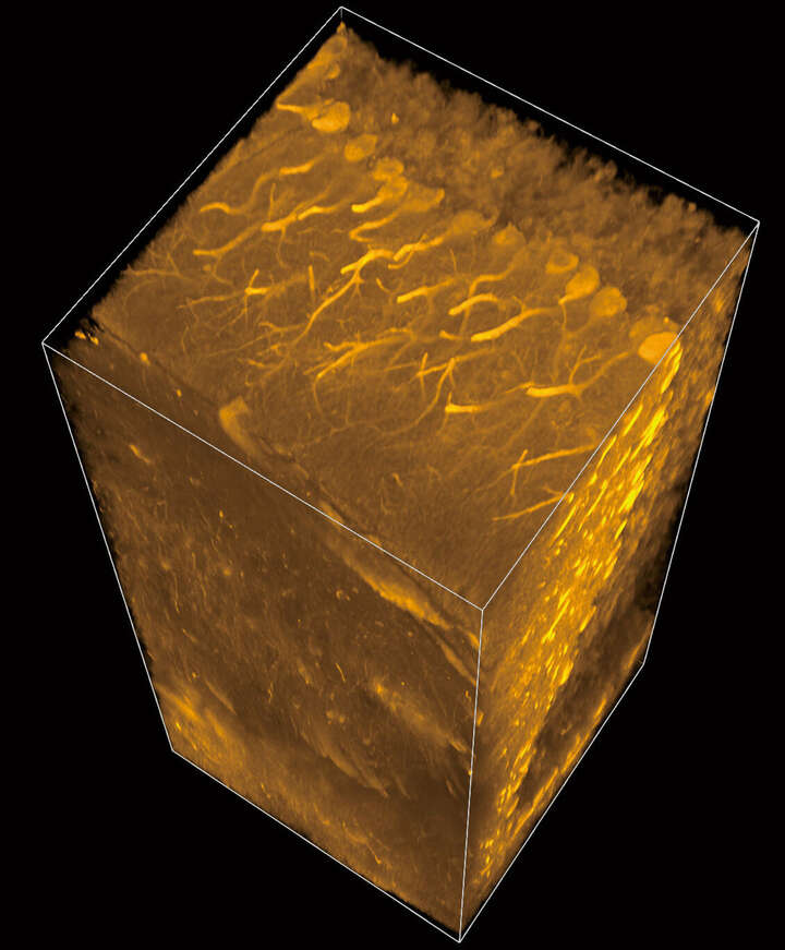
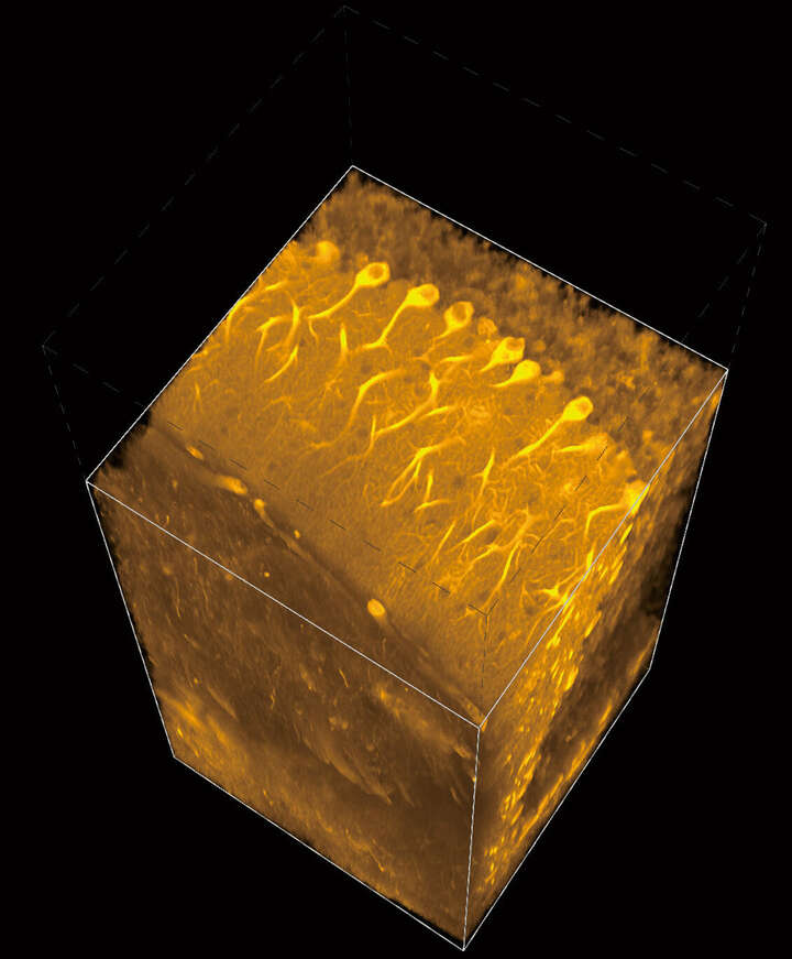
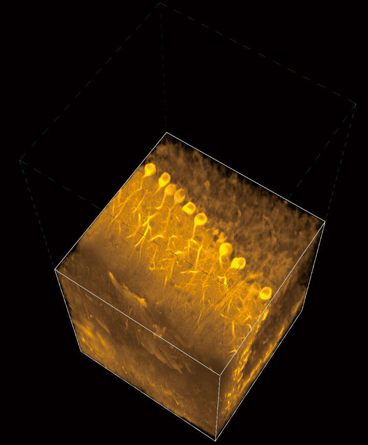
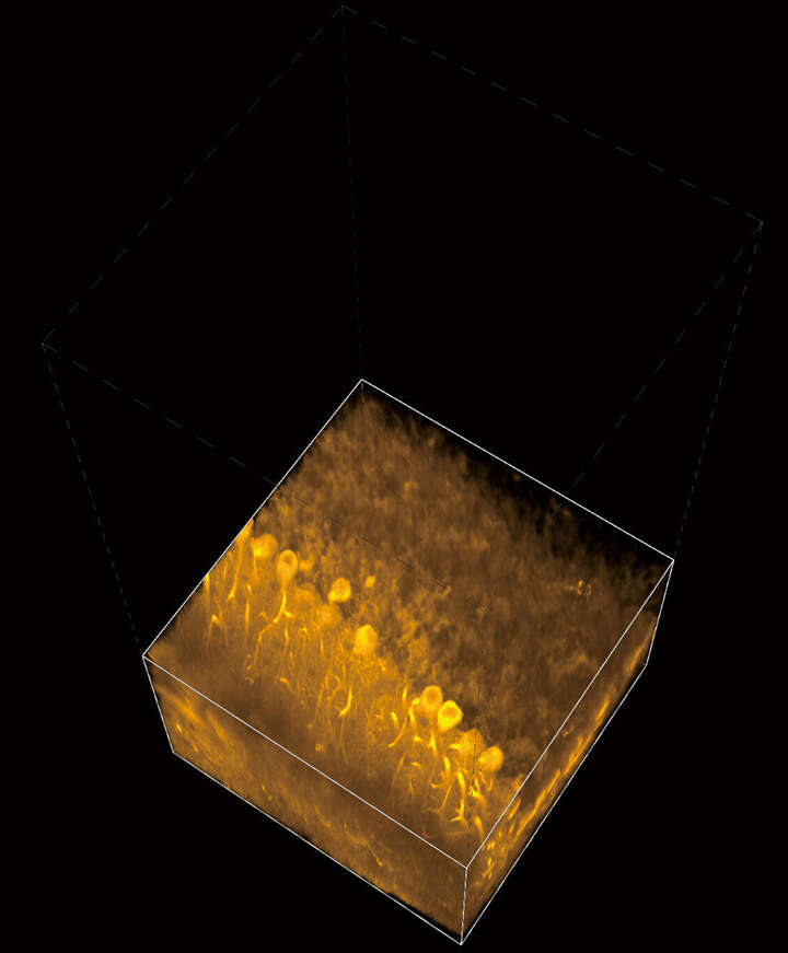
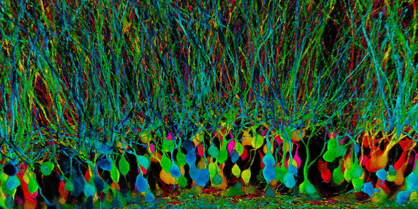
Transgenic Mouse Cerebellum Neurons (0:04)
Cutaway video of Purkinje neurons in the cerebellum of an LC3-GFP transgenic mouse acquired with a CFI75 Apochromat LWD 20XC W objective lens and deconvolved using the NIS-Elements software.
Courtesy of Dr. L. Dubreil, Dr. J. Pichon and Pr MA Colle, CENN at PAnTher UMR703 INRAE/Oniris, Nantes France
Amazingly sensitive
The AX R MP maximizes signal collection by placing detectors as close to the sample as possible. Sensitivity is improved by utilizing the latest electrical and detector designs to drastically reduce noise that normally obscures dim signals. Detector configurations are flexible allowing the system to be tailored to any experimental needs.
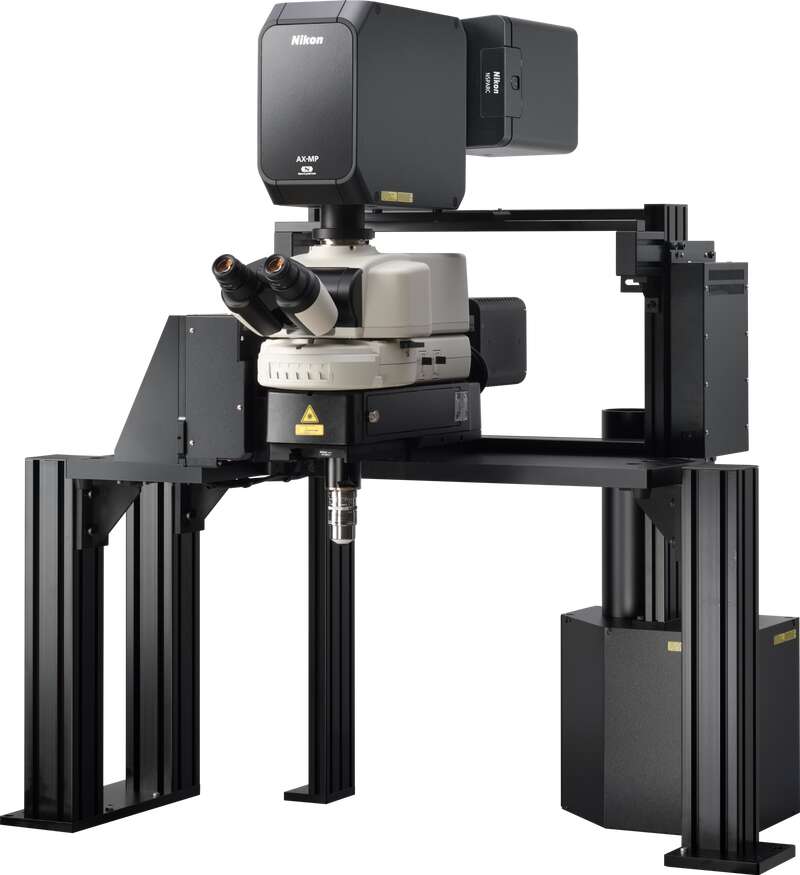
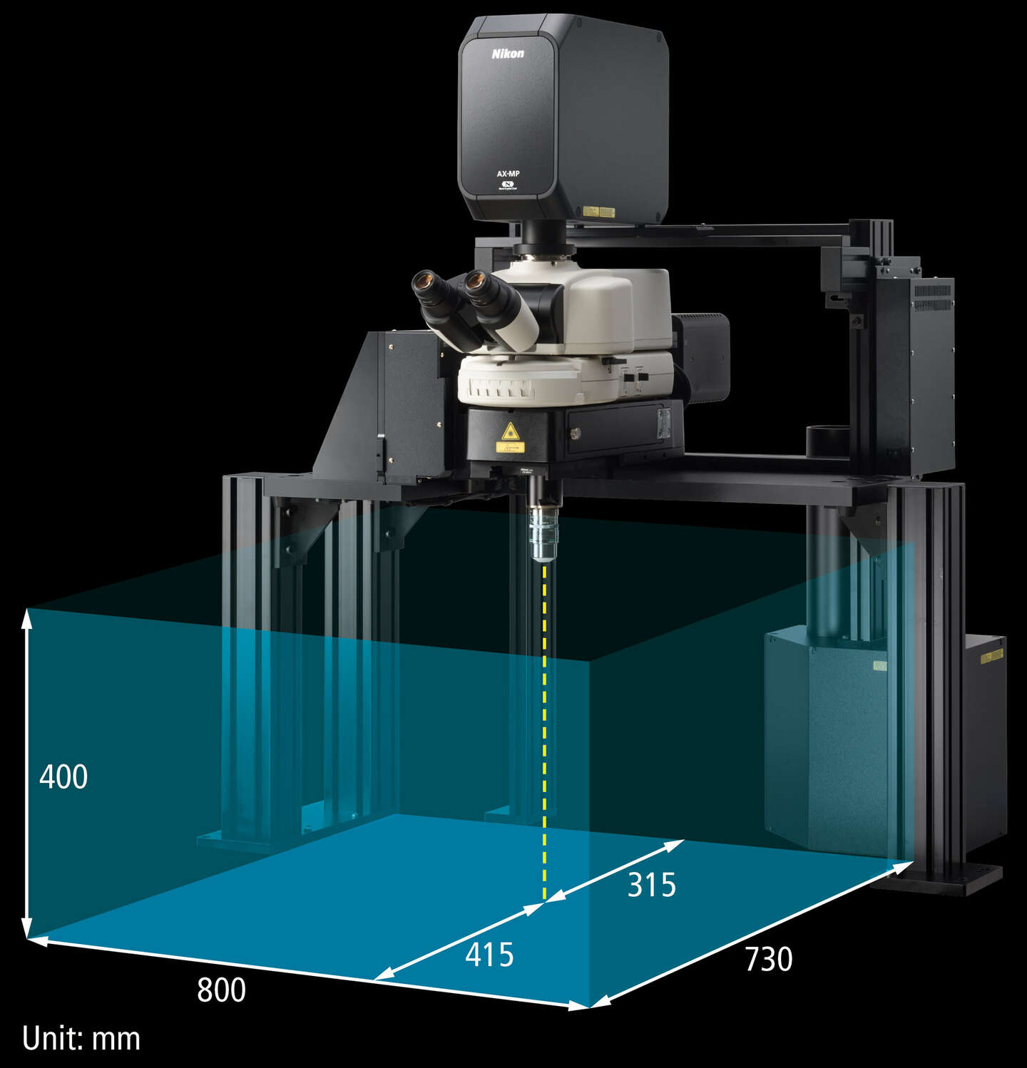
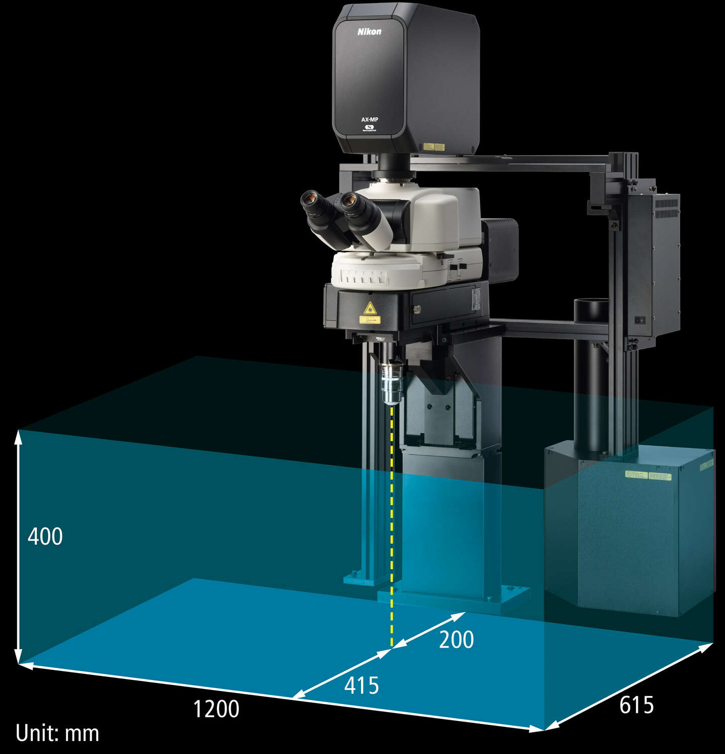
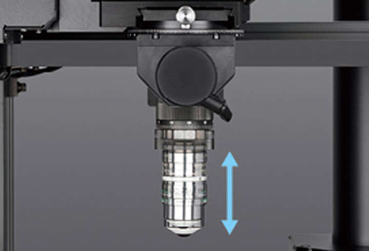
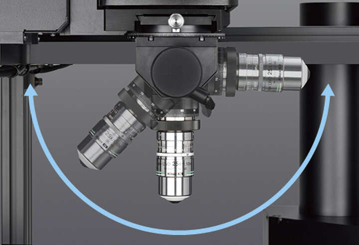
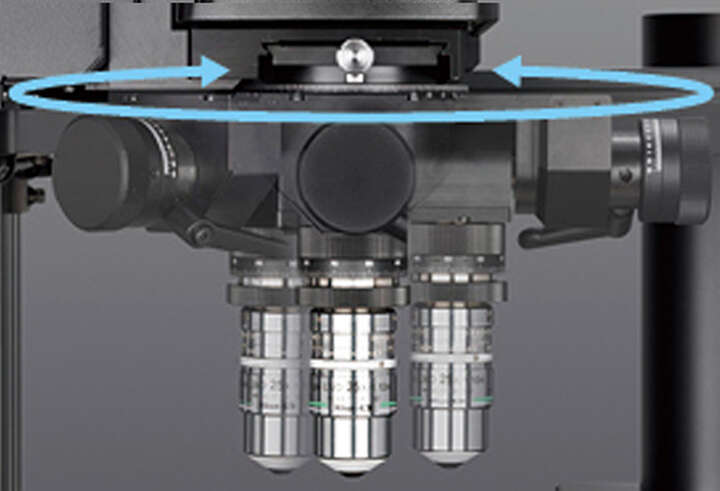
Space to expand
The AX-FN series of microscopes prioritizes flexibility and free space under the objective. This means experimental design is limited only by the users’ imagination, rather than the body of the microscope.
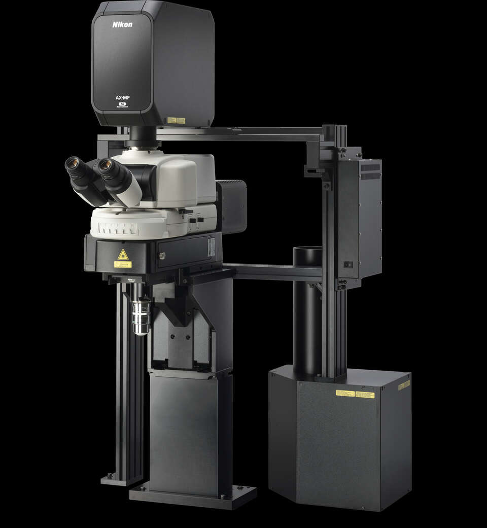
For systems requiring depth
For systems requiring breadth
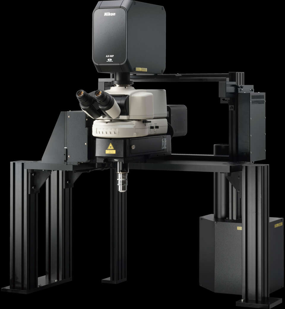
The motorized upright microscope dedicated for AX R MP provides up to 40 cm under the objective in two unique configurations. Both stands provide a large amount of free space around the sample improving positioning, flexibility and accessibility.
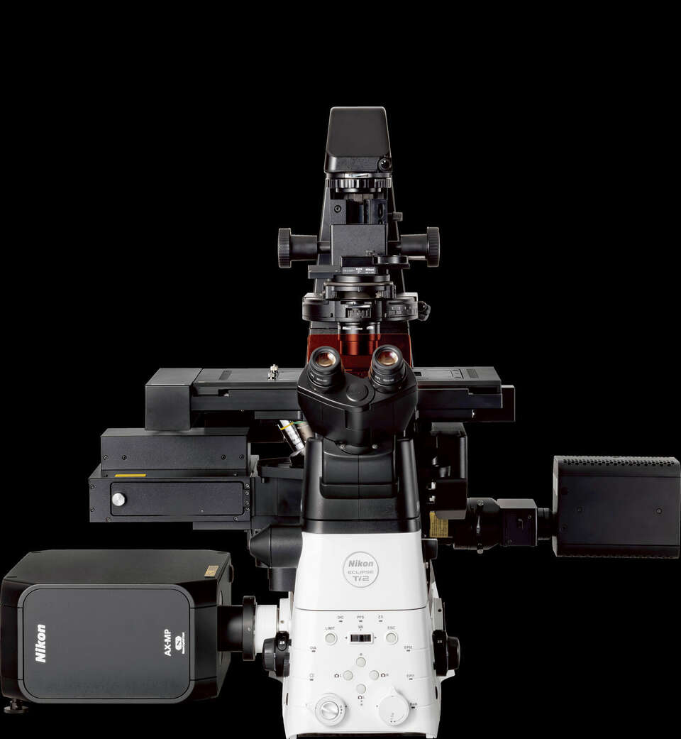
A system built for intravital microscopy
The AX R MP is designed from top to bottom to deliver maximum performance and flexibility for intravital imaging. Built to be fast, without sacrificing image quality, this system delivers reliable data in even the most difficult conditions. With unique AX-FN microscope stands, space is prioritized allowing access to new experimental designs and samples. Auto-alignment brings day-to-day reliability that is absolutely essential when precious intravital data is being collected.
Accommodate any sample
Experiments are best performed with minimal disturbance to the sample. Utilizing Nikon’s new CFI75 tilting nosepiece, the objective can be oriented to image samples in their natural positions, minimizing stress and uncontrolled variables. This nosepiece is compatible with Piezo Z, maximizing 3D image speed in these challenging samples.
Piezo Z device stroke: 450 μm
Support for visible light imaging
The AX R MP supports observation not only at infrared wavelengths, but also at visible wavelengths. It enables both multiphoton imaging and confocal imaging with a single microscope. It also enables simultaneous photostimulation and imaging using two different wavelengths.
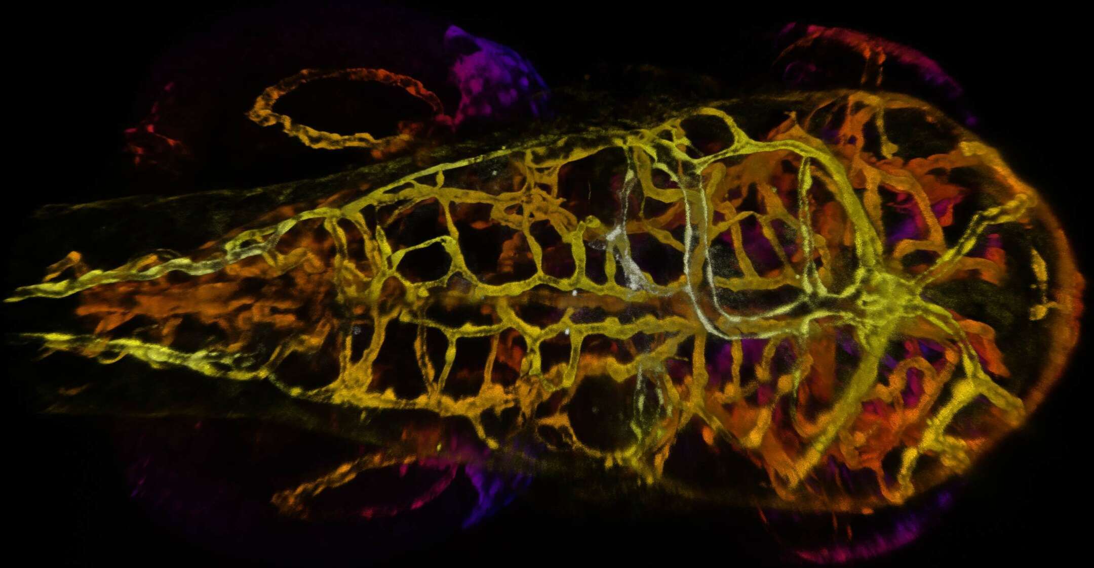
Blood vessels in an embryonic zebrafish labeled with GFP and RFP. Imaged using a CFI75 Apochromat LWD 20XC W objective lens.
Courtesy of Erika Dreikorn and Dr. Beth Roman, Department of Human Genetics, University of Pittsburgh Graduate School of Public Health
Opti-Microscan photostimulator (optional)
Photostimulation using wavelengths* of 400 nm, 488 nm, and 561 nm enables simultaneous visible light stimulation and IR imaging. Stimulation modes include simultaneous, sequential, and manual stimulation.
* Limited by the specifications of the filter cube used.
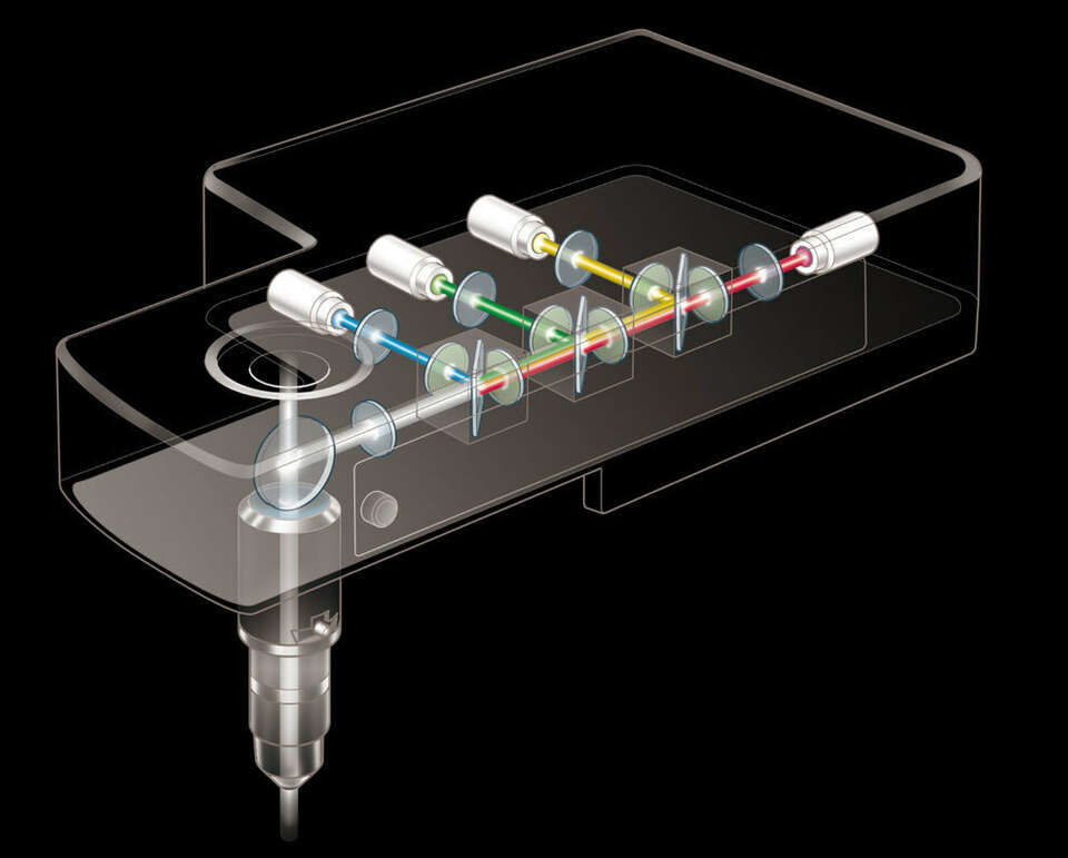
DUX-VB high-sensitivity visible light detector unit
The transmission wavelength band of the LVF (Linear Variable Filter) employed in the DUX-VB gradually changes depending on its location, enabling continuous tuning of the wavelength detection setting within a range of 400 nm to 750 nm. From 2 to 4 channels can be selected, and high sensitivity GaAsP PMT can be used for all channels.
Specifications
| Main body | AX-FNSP | AX-FNGP |
|---|---|---|
| Optical system | Infinity optical system | |
| Microscope stands | AX-FNSP Single Stand | AX-FNGP Gate Stand |
| Focusing | AX-FN Focusing Nosepiece Unit Motorized coaxial coarse/fine focusing Focusing stroke: Up 13 mm/Down 2 mm*1, *2, Minimum step: 0.02 μm, Motorized escape and refocus mechanism Focal plane: 400 mm above the surface of the vibration isolated table |
|
| Controls | AX-FNCTL Control Box AX-FNHC Hub Controller (For controlling Focusing Nosepiece Unit, Diascopic Illumination System, Stage Joystick, Motorized Epi-fluorescence Cube Turret, Motorized Quadrocular Tilting Tube 2 and DSC Zooming Port) |
|
| Stages | AX-FNSP | AX-FNGP |
|---|---|---|
| Adapter | AX-FNSA Stage Adapter, supporting both manual and motorized XY stages. Stage height: adjustable to 2 positions depending on sample size (400 mm/200 mm from the surface of the vibration isolated table) | |
| Stage | FN-3PS2 XY stage, Cross travel 29.5 (X) x 29.5 (Y) mm, with 2 auxiliary plates AX-FNS-E Motorized XY stage, Cross travel ±34 (X) x ±27 (Y) mm |
|
| Epi-fluorescent illuminator | AX-FNSP | AX-FNGP |
|---|---|---|
| Illumination unit | NI-FLEI-2 Epi-fluorescence attachment | |
| Light source | D-LEDI Fluorescent LED Illumination System | |
| Filter cube turret | 6 mountable filter cubes, shutter function
|
|
| Photostimulation device | AX-FNBPU Stimulation Back Port, 6 mountable filter cubes, Fluorescence imaging and simultaneous stimulation imaging can be switched | |
| Diascopic illuminator | AX-FNSP | AX-FNGP |
|---|---|---|
| Illumination unit | AX-FNDIA Diascopic Unit 4 filter slider attachable, Condenser holder stroke: Up 2.5 mm/Down 1.8 mm, NI-PT Polarizer Turret mountable |
— |
| Light source | C-LL High Color Rendering LED Lamphouse | — |
| Shutter | NI-SH-E Motorized Shutter | — |
| Condenser | FN-C LWD condenser, O.D. 8.2 mm, NA: 0.78 | — |
| Polarizer Turret | NI-PT Polarizer Turret, Visible or infrared polarizer attachable | — |
| AX-FNSP | AX-FNGP | |
|---|---|---|
| Tubes | Pupillary distance: 50-75 mm, Inclination angle: 15-35 degrees, Eyepiece/Upper port/Rear port: 100/0/0, 0/100/0, 0/0/100 via DSC Zooming Port
|
|
| Eyepieces (F.O.V. (mm)) | CFI 10X (22), CFI 12.5X (16), CFI 15X (14.5), CFI UW 10X (25) | |
| Photodetector | AX-NEU Non-descanned EPI Upright Detector | |
| Nosepieces |
|
|
| Observation methods | Brightfield, Epi-fluorescence, DIC | |
| Power consumption | 100W | |
| Weight (approx.) | 66 kg (fully motorized fluorescence system, with diascopic illuminator) | 66 kg (fully motorized fluorescence system) |
| Scan head | AX R MP |
|---|---|
| Type | AX-SHRM AX R MP Scan Head & Controller |
| FOV | φ22 mm |
| Standard image acquisition | Galvano scanner Pixel size: max. 8192 x 8192 pixels Scanning speed: max. 240 fps (512 x 16 pixels), 10 fps (512 x 512 pixels) |
| High-speed image acquisition | Resonant scanner Pixel size: max. 2048 x 2048 pixels Scanning speed: max. 720 fps (2048 x 16 pixels for 2K, 1024 x 16 pixels for 1K), 30 fps (2048 x 512 pixels for 2K, 1024 x 512 pixels for 1K) |
| Scan mode | Line scanning, bi-direction scanning and averaging |
| Simultaneous acquisition | Max. 5 channels (including a diascopic detector channel) |
| IR laser wavelength range | 700-1080 nm (1080 system), 820-1300 nm (1300 system) |
| Dichroic mirror | Position: 6 |
| Pinhole | 6-153 µm variable |
| Zoom | 1–1000X continuously variable |
| Input/output port | 2 laser input ports 2 signal output ports |
| Laser for multiphoton microscopy | AX R MP |
|---|---|
| Single 1080 system | Mai Tai HP/eHP DeepSee, Chameleon Vision II, Axon 920*6,Chameleon Discovery LX |
| Dual 1080 system | Chameleon Vision II + Axon 920*6, Axon 920*6 + Axon 1064*7, Chameleon Discovery LX + Axon 920*6 |
| Single 1300 system | InSight X3 + InSight X3+, Chameleon Discovery NX |
| Dual 1300 system | InSight X3 Dual Option, InSight X3+ Dual Option, Chameleon Discovery NX, Chameleon Discovery NX + Axon 920*6 |
| Incident optics | 700-1080 nm (1080 system), 820-1300 nm (1300 system), auto alignment |
| Modulation | Method: AOM (Acousto-Optic Modulator) device Control: power control, ROI exposure control |
| Laser for confocal microscopy (option) | AX R MP |
|---|---|
| 4-laser unit | 405 nm, 488 nm, 561 nm and 640 nm lasers are installed |
| 5-laser unit | 405 nm, 488 nm, 561 nm, 594 nm and 640 nm lasers are installed |
| 6-laser unit | 405 nm, 445 nm, 488 nm, 515 nm, 561 nm and 640 nm lasers are installed |
| NDD for multiphoton microscopy | AX R MP |
|---|---|
| NDD EPI unit AX-NEI (for Ti2-E) and AX-NEU (for AX-FNSP/FNGP) | Detectable wavelength range: 400-650 nm (1080 system), 400-750 nm (1300 system)
Detectors: 2 GaAsP PMTs (4 GaAsP PMTs, or 3 GaAsP PMTs + 1 multi-alkali PMT are possible by adding options) |
| Visible stimulation/IR imaging (option) | AX R MP |
|---|---|
| Opti-Microscan Photostimulator (for AX-FNSP/AX-FNGP) | Stimulation wavelength: 405 nm, 488 nm, 561 nm; Excitation wavelength for imaging: 800-1080 nm (1080 system), 820-1080 nm (1300 system) Stimulation speed: Max. 1 ms (point stimulation), Max. 20 μs/pixel (ROI stimulation) Stimulation modes: simultaneous, sequential, manual Stimulation area: square inscribed within a 22 mm-diameter circle, stimulation ROI: arbitrary pattern, no number limit |
| Diascopic detector (option) | AX R MP |
|---|---|
| AX-DUT-MP (for AX-FNSP/Ti2-E)*8 | Detectable wavelength range: 400-920 nm Detector: Multi-alkali PMT |
| Detector for confocal / multiphoton microscopy (option) | AX R MP |
|---|---|
| DUX-VB detector unit | Detectable wavelength range: 400-650 nm (with IR laser), 400-750 nm (with visible laser); Detection width: 10 nm to 320 nm Maximum pixel size: 8192 x 8192 (with galvano scanner) Wavelength resolution: 5 nm, wavelength range variable in 1 nm steps Compatible with galvano and resonant scanners2 or 4 channels (Multi-alkali PMT or GaAsP PMT options) |
| DUX-ST detector unit*9 | Detectable wavelength range: 400-750 nm 2 or 4 channels (Multi-alkali PMT or GaAsP PMT options) |
| NSPARC Detector Unit | Equipped with SPPC (Single Pixel Photon Counter) array detector Up to 7 barrier filters can be mounted (Mountable filter: QuadBand446/523/600/677, 452/45, 525/50, 593/46, 700/75) With galvano scanner: Can be used with X resolution of 64 to 8192 pixels, Y resolution of 2 to 8192 pixels With resonant scanner: Can be used with X resolution of 256, 512, 1024 and 2048 pixels, Y resolution of 128 to 2048 pixels |
| Option | AX R MP |
|---|---|
| Motorized XYZ | Motorized XY stage (for AX-FNSP/FNGP/Ti2-E), High-speed piezo Z stage (for Ti2-E), High-speed piezo objective-positioning system (for AX-FNSP/FNGP) |
| Nosepiece for AX-FNSP/FNGP | AX-FNTN-H CFI75 single tilting nosepiece*4 |
| Software | AX R MP |
|---|---|
| Acquisition/analysis | Imaging software (equipped with Denoise.ai noise reduction function): NIS-Elements C or NIS-Elements C-ER |
| Display/image generation | 2D analysis, 3D volume rendering/orthogonal, 4D analysis, spectral unmixing |
| Image format | JP2, JPG, TIFF, BMP, GIF, PNG, ND2, JFF, JTF, AVI, ICS/IDS |
| Application | FRAP, FLIP, FRET(option), photoactivation, 3D time-lapse imaging, multipoint time-lapse imaging, colocalization |
| Control computer | AX R MP |
|---|---|
| OS | Windows®10 Pro 64 bit, Microsoft Windows® 11 Pro*10 |
| AX R MP | |
|---|---|
| Visible stimulation/IR imaging (option) | Stimulation wavelength: 405 nm, 488 nm, 561 nm Excitation wavelength for Imaging: 800-1080 nm (1080 system), 820-1080 nm (1300 system) |
| Compatible microscopes | Dedicated AX-FNSP/AX-GNGP motorized upright microscope system, ECLIPSE Ti2-E motorized inverted microscope |
| Z step | AX-FNSP/FNGP: 0.02 µm, Ti2-E: 0.02 µm |
| Recommended installation conditions | Temperature 20 – 25˚C, ± 1˚C, air conditioning at all hours Humidity 60% RH or less (no condensation) |
*1 Based on the focus position
*2 Software controlled value
*3 DIC prism slider cannot be attached
*4 FOV 12, Usable objectives: CFI75 LWD 16X W, CFI75 Apochromat LWD 20XC W, CFI75 Apochromat 25XC W, CFI75 Apochromat 25XC W 1300
*5 Cannot be used with diascopic illumination. The FN-MN-H cannot be used with diascopic illumination only when the 400 μm objective piezo positioner (PI) is attached.
*6 Axon 920 : Axon 920-1、Axon 920-2
*7 Axon 1064 : Axon 1064-1、Axon 1064-3
*8 Cannot be mounted on AX-FNGP
*9 Must be used with a confocal laser.
*10 Windows 10 and Windows 11 are trademarks or registered trademarks of Microsoft Corporation in the United States and other countries.