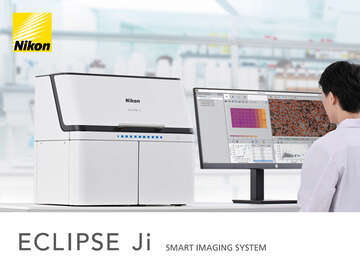

Overview & Features
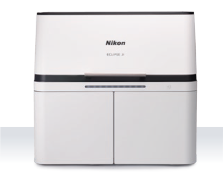
Research Microscope Power in a Benchtop Assay Instrument
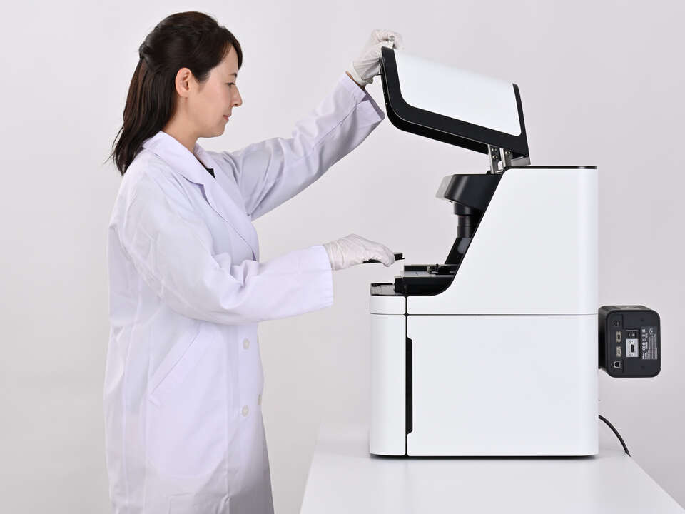
Simple Operation
Minimal experimental setup and complexity by AI-driven assays and analysis.
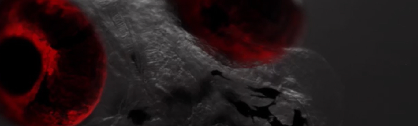
Easy Cellular Imaging
No need to master complicated microscope hardware and software – ECLIPSE Ji makes data collection and cellular imaging assays easy.
High Quality Optics
Renowned Nikon optics provide clear and sharp images on plate assay devices.
Features
Smart Experiments with Automated Assays
Utilizing Nikon’s precision optical hardware, all of the advantages of high sensitivity and resolution from a research-level microscope are retained by an AI-driven, easy-to-operate benchtop laboratory instrument.
Preconfigured and optimized turnkey assay experiments minimize time spent defining parameters, maximizing data collection.
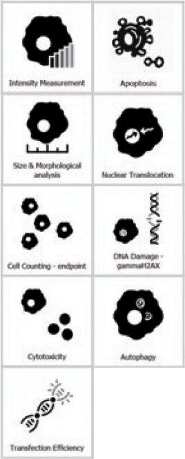
Standard Assays
Intensity Measurement
Compares protein expression level changes in cells and cell nuclei in multiple wells.
Cell Counting (endpoint)
Counts the number of cell nuclei in a fixed sample and the area of the well occupied by cells.
Cell Counting (proliferation)
Measure changes in confluency and count nuclei in live samples over time.
Transfection Efficiency
Investigates the percentage of cells expressing the target protein, and reports the expression efficiency.
Size & Morphological analysis
Analyzes cellular morphology with measurements of the cell nucleus, cytoplasm, and the size of the cell region.
Cytotoxicity
Measures the percentage of dead cells among all cells and evaluates cytotoxicity.
Optional Assays
Nuclear Translocation
Measures the nuclear translocation of NF-κB, indicating an extra-cellular stimulus.
Autophagy
Measures the number of autophagosomes, their area, and their fluorescence intensity.
Phagocytosis
Measures the number, area, and fluorescence intensity of bioparticles taken into the cell by phagocytosis.
Endocytosis
Measures the number, area, and fluorescence intensity of granules formed by endocytosis, which are taken up from outside the cell.
Micronucleus test
Measures the number of cells containing micronuclei or multiple nuclei.
Mitochondrial toxicity
Measures the number, area, and fluorescence intensity of mitochondria.
Neurite Outgrowth
Measure the number and length of neurites protruding from neuron cell bodies.
Wound Healing
Measure the recovery of filled area in an artificially-created wound over time.
Cell Cycle (Fucci)
Measure the proportion of cells in each cell cycle phase based on Fucci, the genetically-encoded, fluorescent cell cycle reporter system.
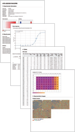
Effortless Results using AI
ECLIPSE Ji’s Smart Experiment software interface uses newly developed artificial intelligence (AI), implemented to minimize errors and maximize data collection.
| Input | Output | ||
|---|---|---|---|
Load a plate |
Select assay / Input basic information |
Check the shooting results and analysis results |
Report |
| step 1 | step 2 | step 3 | step 4 |
| ECLIPSE Ji |
Pre-AcquisitionAI automatically judges plate status and automatically adjusts acquisition conditions |
Actual Acquisition/analysis/data displayAutomatic acquisition, data extraction, graphing under optimized acquisition conditions |
ReportingOne click export of the data |
Single-Cell Imaging and Analysis with Ease
AI based on Deep Learning defines acquisition settings and image analysis parameters, saving researchers valuable time at the microscope.
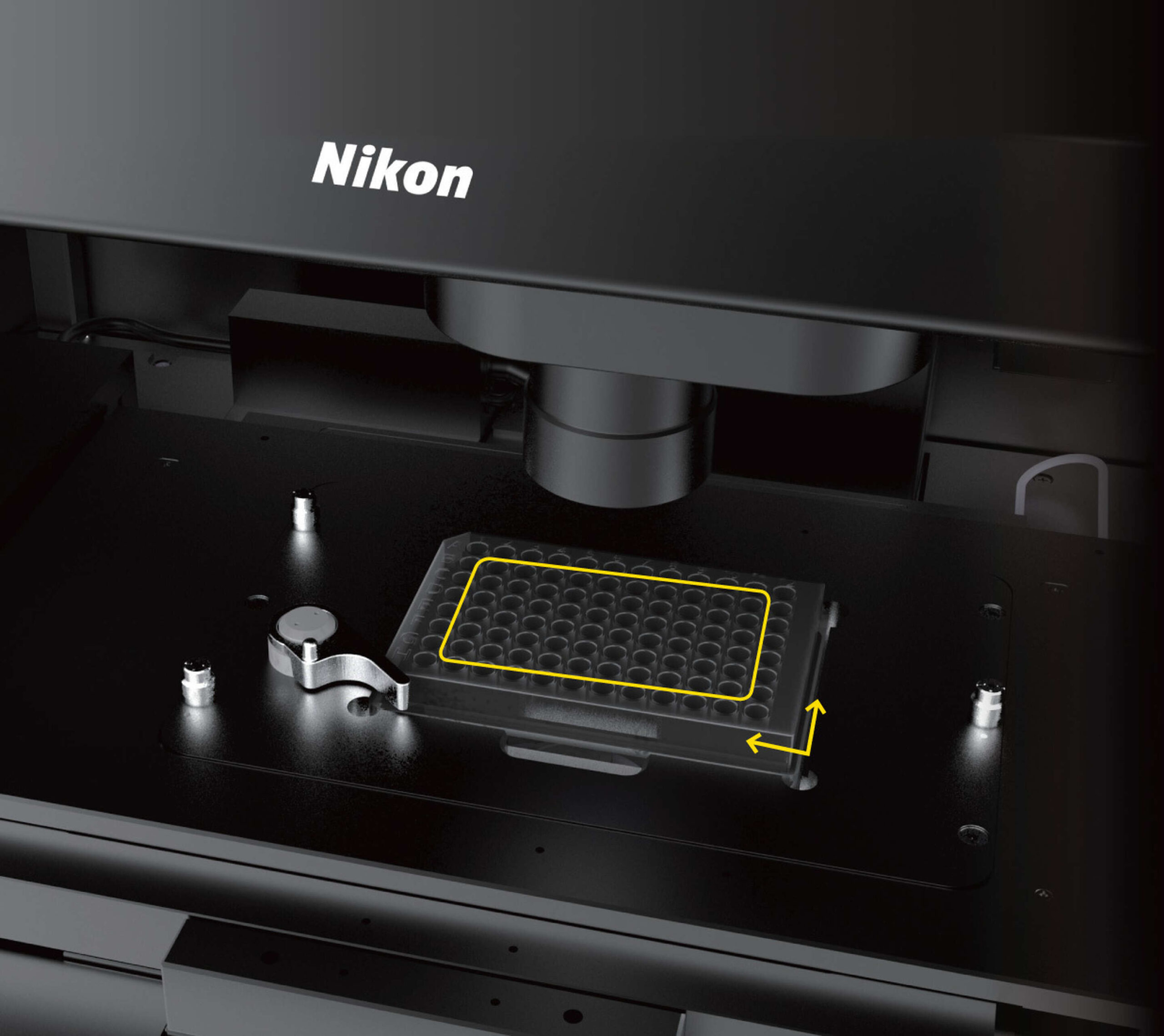
Automatic Plate Detection
Plate type and dimensions are automatically detected. There is no need to select from lists or manually enter plate data.
Automatic detection of samples
A rapid preview across the entire plate determines which wells have sample present, allowing users to easily skip empty wells.
Automatic calculation of optimal exposure settings
There is no need for troublesome tuning of light intensity and exposure time. The optimal exposure settings for image analysis are automatically calculated from the luminance values of all wells.
Automatic alignment of the plate
No alignment work is required. The system automatically detects and corrects the plate position.
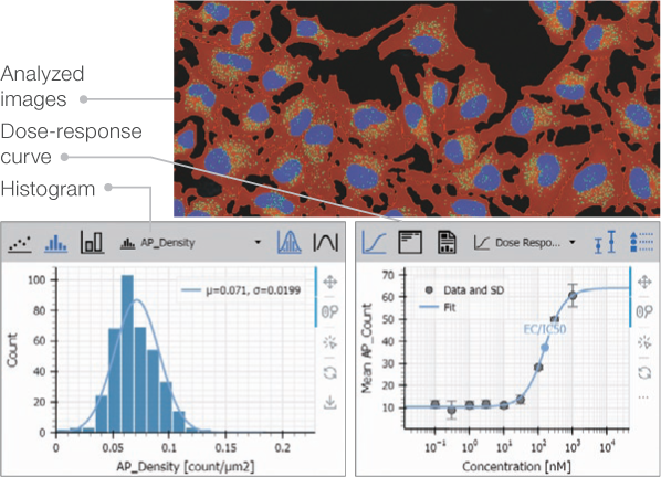
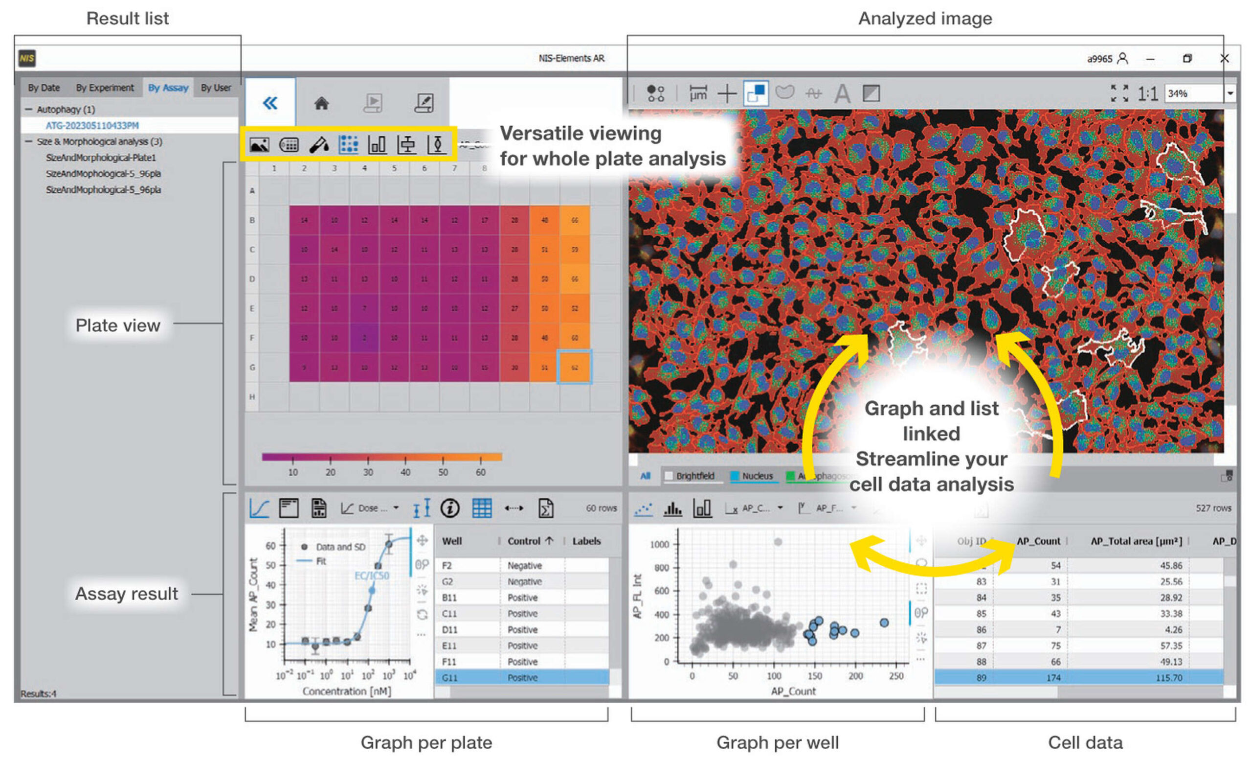
User Interface Designed for Data-Rich Microscopy
Images and corresponding analysis data for the plate, well, and each cell is contained in an interactive and linked interface. Users can navigate and quickly visualize trends and results.
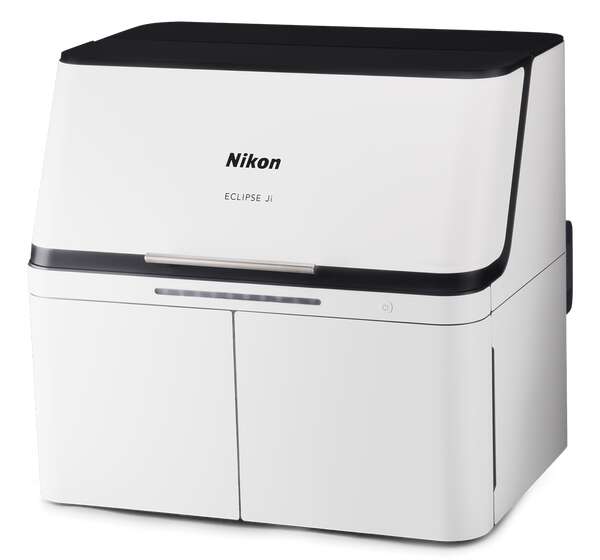
ECLIPSE Ji: a Multi-Role Digital Inverted Microscope
Outside of plate assays, ECLIPSE Ji can also serve as a digital research microscope, and can be integrated with a variety of peripheral hardware ranging from filter wheels through confocal systems such as “AX“, or high-sensitivity cameras.
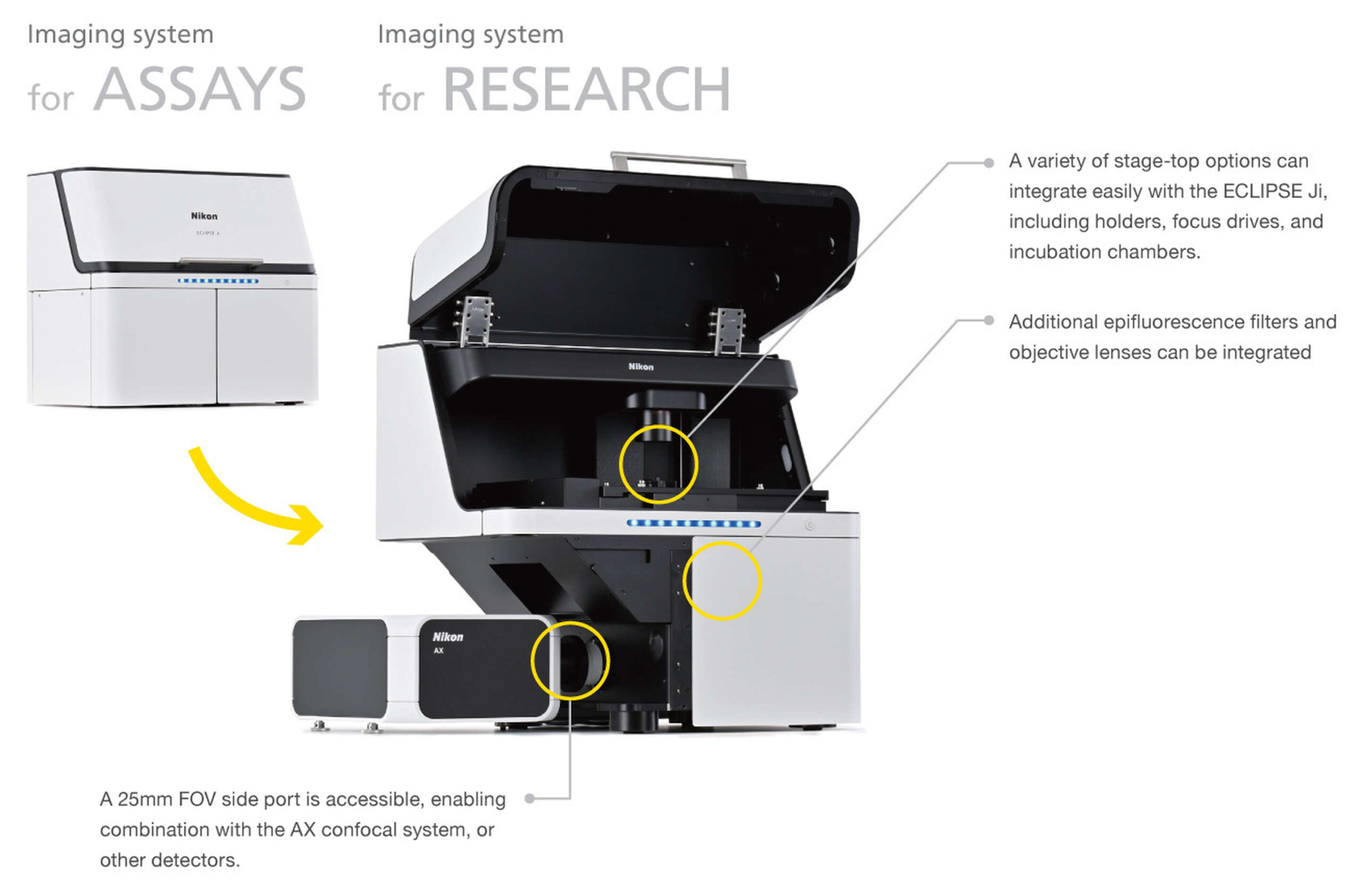
*The design and specifications may differ from the actual product.
Specifications
| ECLIPSE Ji | |
|---|---|
| Observation methods | Brightfield, Epi-fluorescence |
| Optical system | CFI Infinity Optical System Observation Optical System: Inverted image observationFOV 25Optical path switching: Switching between the built-in camera optical system and the left side port |
| Built-in camera | Imaging device: 7.8 megapixel monochrome CMOS sensor Output signal Tone: Monochrome 12 bits/8 bitsFrame rate: Maximum 18 fps Output pixel number: 2800×2800 pixels (when assay used) |
| Focusing | Drive system: Motorized (Via PFS nosepiece objective lens up/down movement)Focusing stroke: About 10 mm Focusing speed: Maximum driving speed 2.5 mm/sec |
| PFS* | Focal point maintenance control: Infrared light projecting method Applicable observation methods: Brightfield, Fluorescence observation |
| Transmission illumination section | Koehler illumination Light source: LED |
| Stage | Stroke: X: ±59 mm, Y: ±39.5 mm Maximum drive speed About 25 mm/sec |
| Nosepiece | Objective lens mounting holes: 6 Nosepiece drive method: Motorized |
| Fluorescence cube turret | Number of filter cubes that can be mounted: 6 (Compatible with wide-field filter cubes) Turret drive method: Motorized |
| Light distribution section | Light source used: D-LEDI2 fluoresce LED light source |
| PC interface | USB interface: Device interface (for built-in camera) B connector USB 3.0 (SuperSpeed) |
| Input rating | 100V-240VAC±10%, 3.0 A, 50/60 Hz |
| Power consumption | 320 W |
| Power source cord | – 100 to 120 V: Power source cord of 3 conductor grounding Type SVT, NO.18 AWG, 3 m long maximum, rated at 125VAC minimum with detachable receptacles conforming to UL specifications – 220 to 240 V: Power source cord of 3 conductor grounding Type H05VV-F 1 mm2, 3 m long maximum, rated at 250VAC minimum with detachable receptacles conforming to EU/EN specifications |
*PFS: a function that automatically corrects focal point displacement due to the passage of time and/or stage movement.