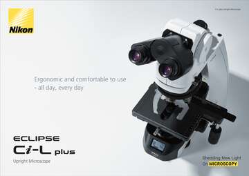

Overview & Features
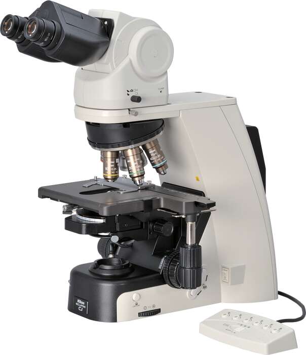
ECLIPSE Ci Series-To meet the demands of clinical laboratory specialists and researchers, Nikon has reviewed all aspects of microscope usability to develop the ECLIPSE Ci series of microscopes, which combines superior functionality with operational ease. The Ci is designed to ensure natural posture while viewing images, sample changing and capturing images. Bright, eco-friendly LED illumination reduces the need for frequent lamp replacement*. A variety of accessories are available that support various imaging techniques.
* Halogen model (Ci-S) is also available
This product is available for clinical use in the United States. Please contact your local Nikon dealer for information on clinical use of this product in your country.
Features
To meet the demands of clinical laboratory specialists and researchers, Nikon has reviewed all aspects of microscope usability to develop the ECLIPSE Ci series of microscopes, which combines superior functionality with operational ease. The Ci is designed to ensure natural posture while viewing images, sample changing and capturing images. Bright, eco-friendly LED illumination reduces the need for frequent lamp replacement*. A variety of accessories are available that support various imaging techniques.
* Halogen model (Ci-S) is also available
This product is available for clinical use in the United States. Please contact your local Nikon dealer for information on clinical use of this product in your country.
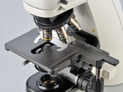
Motorized magnification switching
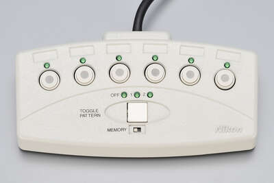
Magnification can be easily changed during observation with the touch of a button on the ECLIPSE Ci-E microscope body. The buttons can also be programmed for specific objective lenses for easy switching between specific pairs. User-defined light intensity for each magnification is automatically saved and reproduced when magnification is changed. The remote control pad can also be used to easily change magnifications.
Bright and uniform Eco-illumination
The Eco-illumination* is a low-power-consuming, eco-friendly illumination system that produces uniform brightness and reduces the cost and effort of lamp replacement, thanks to its 60,000 hour, high-luminescent LED. By combining a collimator lens, fly-eye optics and LED illumination, bright and edge-to-edge uniform images can be obtained even at high magnifications. The LED illuminator features low-heat generation and provides the same color temperature at every magnification.
* Halogen model (Ci-S) is also available

Enhanced operational ease
Observation with a natural posture
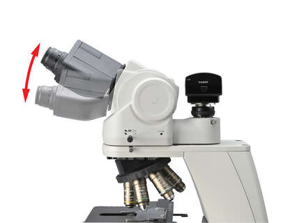
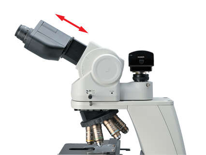
Using the ergonomic binocular tube, which features an eyepiece that can be inclined from 10° to 30°and extended up to 40 mm, the microscope can be adjusted for natural posture. The eyelevel riser lifts the eyepiece tube in 25 mm increments (up to 100 mm*) and can easily be adapted to suit users with different eye-point heights.
* Up to 50 mm with ergonomic binocular tube.
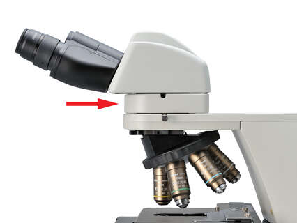
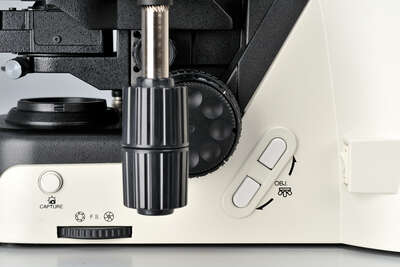
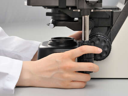
User-friendly stage operation
With the addition of a nosepiece spacer, the stage height can be lowered 20 mm from the standard position, reducing strain during frequent specimen change. The stage handle height can also be changed to ensure a comfortable hand position. The stage height can be locked using the refocusing knob, allowing quick refocusing after specimen changes. The stage is coated with a high-durability, scratch-resistant ceramic coating.
Easy on the eyes, even during extended use
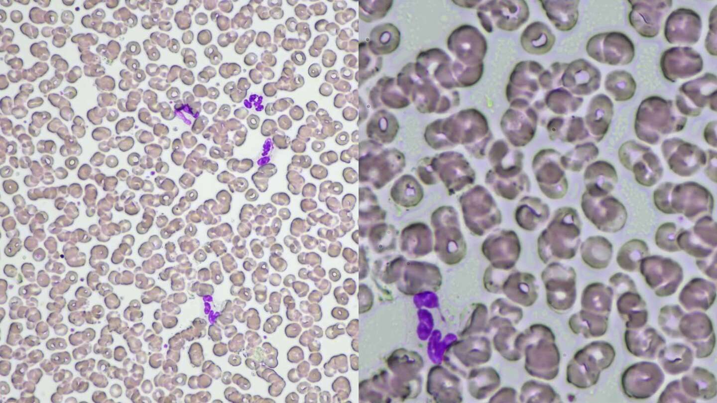
Maintains optimal brightness
The light intensity management (LIM) function developed by Nikon automatically stores any changes to brightness settings. This helps avoid drastic changes in brightness when switching between different magnifications during observation, thereby helping to mitigate eye strain.
| Low-magnification objective lens | High-magnification objective lens |
with LIM
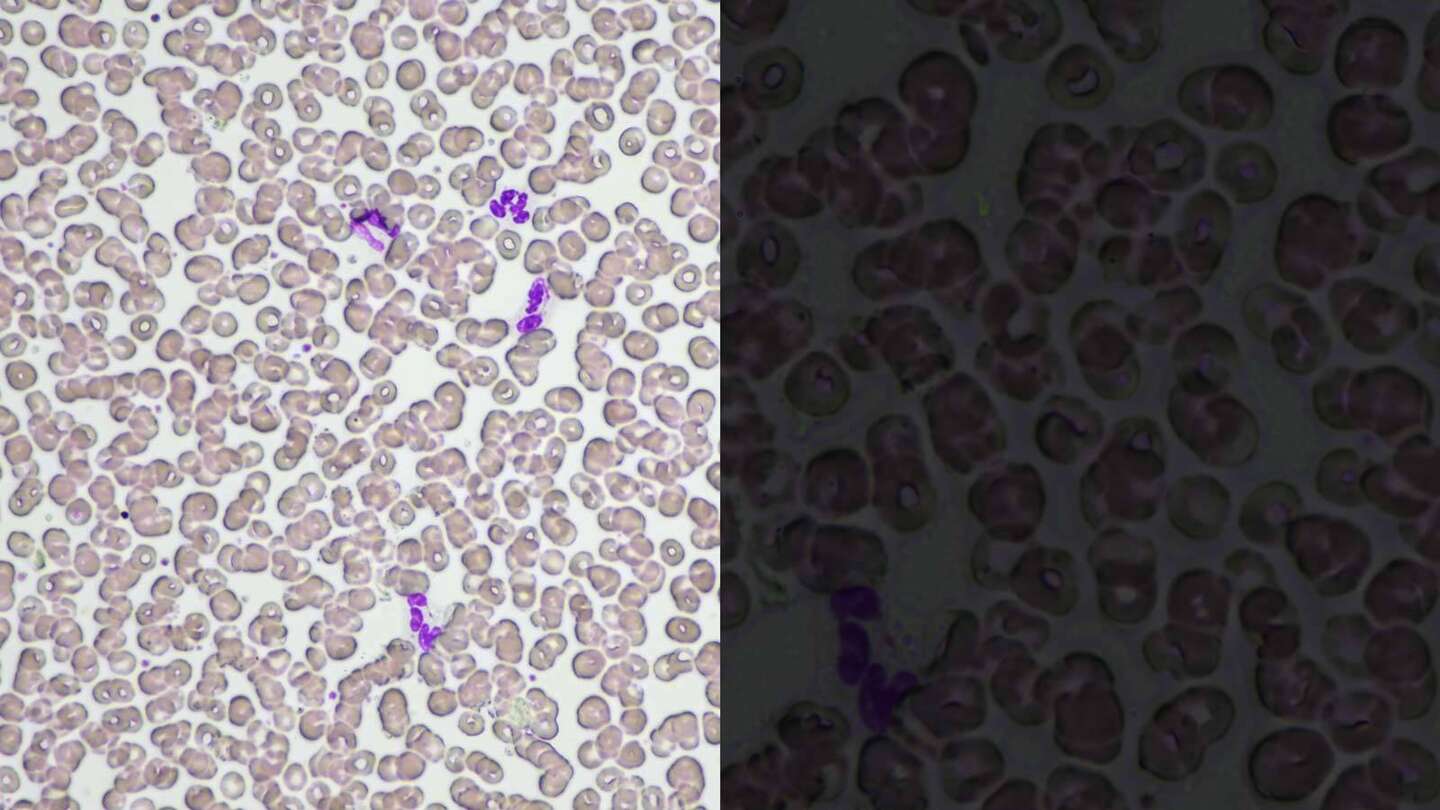
| Low-magnification objective lens | High-magnification objective lens |
without LIM
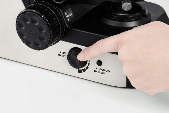
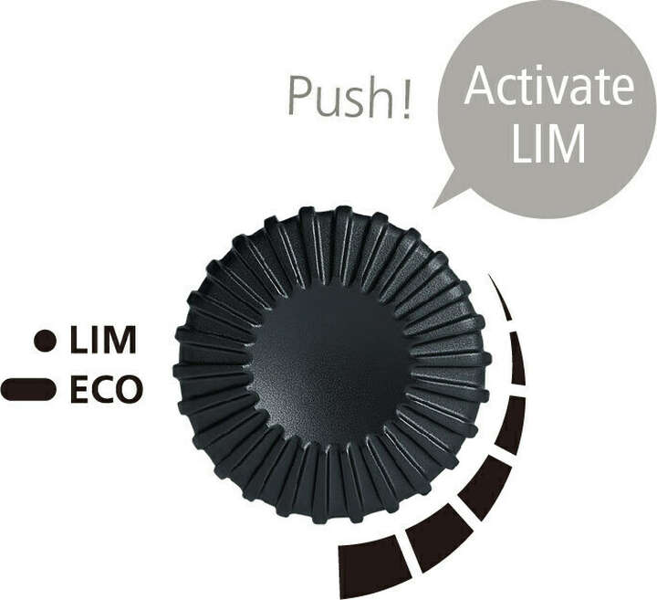
Keep the same observation posture with automatic brightness adjustment
The LIM function saves and recalls the optimal brightness level for each objective, eliminating the need to manually adjust the illuminator and change posture every time you switch objective lenses.
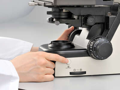
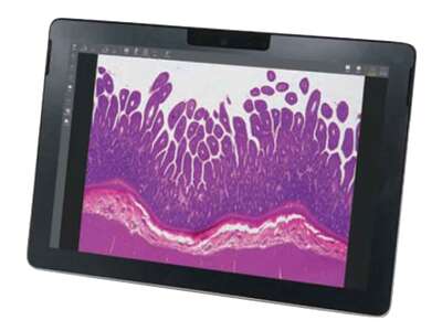
Effortless image capturing
One simple click of the image capture button on the microscope base during observation enables the Digital Sight camera to capture the specimen image. Camera control software for tablet PC NIS-Elements L is equipped with a scene mode that can automatically set the optimum shooting conditions for each observation method. Since it has a network function, you can share images with a remote PC.
* NIS-Elements L is not for clinical diagnostic use.

Enjoy enhanced efficiency during observations
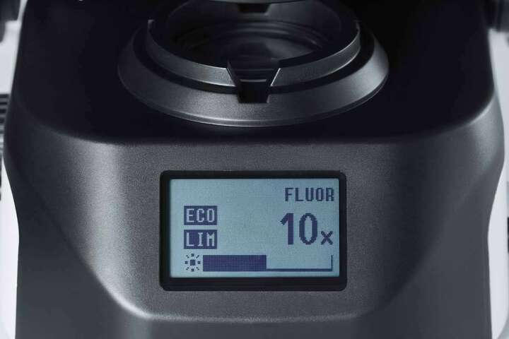
❷ LIM function: ON
❸ Objective name
❹ Magnification
❺ Brightness state
Status Display for easy confirmation at a glance
Quickly and easily check magnification and brightness settings using the display at the base of the microscope without changing your observation posture.
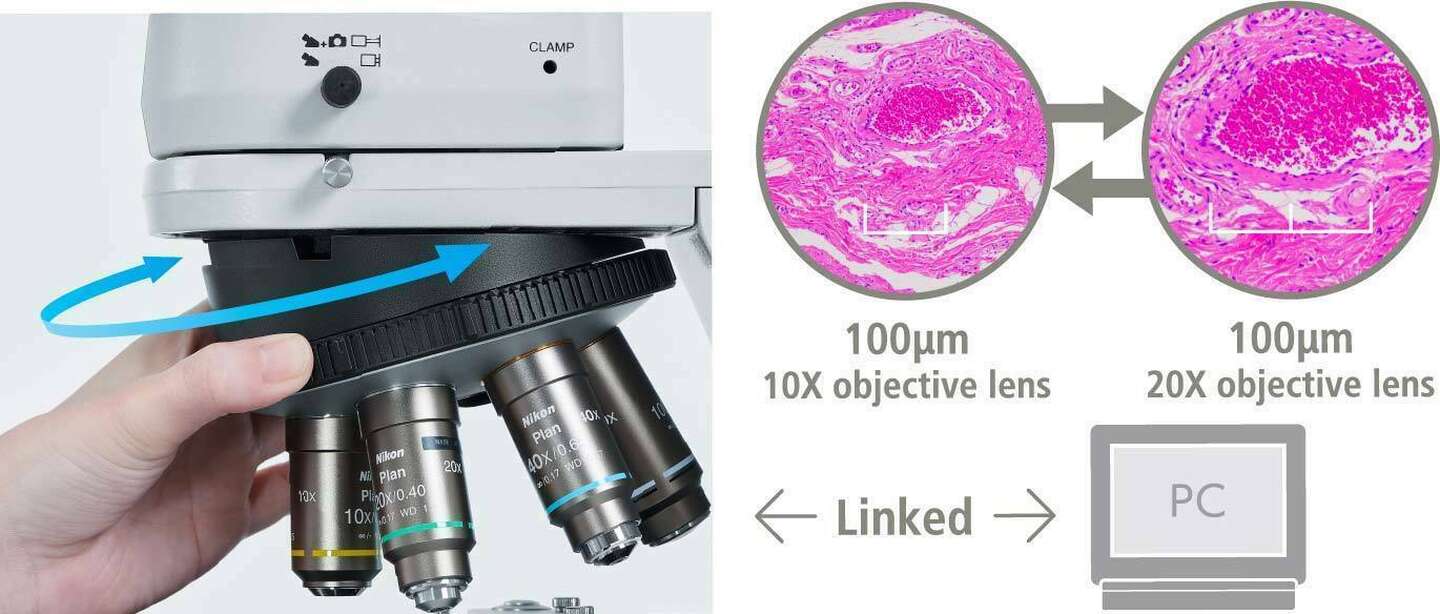
Turning the nosepiece automatically adjusts the scale bar
The scale bar on the PC display changes automatically to match the magnification level, eliminating the need to set the scale manually.
*Sold separately: Software (NIS-Elements D / BR / AR), Nikon recommended camera required
Versatile observation techniques
Phase contrast
High-contrast images with neutral background coloration regardless of the magnification range can be captured. This observation technique is suitable for observation of unstained structures.
Simple polarizing
Ideal for observing bi-refringent samples such as collagen, amyloids and crystals.
* Two types of analyzer are available: intermediate tube type and nosepiece slider type.
Department of Clinical laboratory, Nihon University Itabashi Hospital
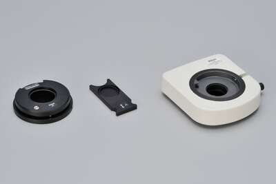
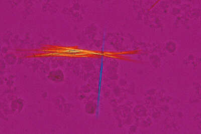
Sensitive color polarizing
Enables Identification of uric acid crystals by changes in the interference color. This technique is ideal for gout and pseudo-gout tests.
* Two types of analyzer are available: intermediate tube type and nosepiece slider type.
Department of Clinical laboratory, Nihon University Itabashi Hospital
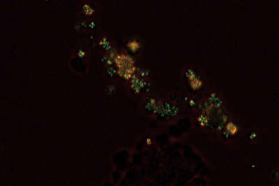
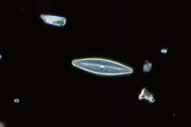
Darkfield
Enables clear observation of blood or minute structures such as flagella. Dry- and oil-type condensers are available. An expander lens is utilized for brighter imaging.
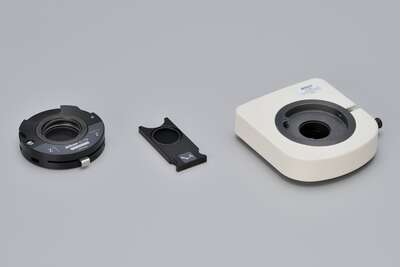
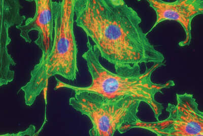
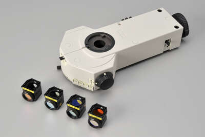
Epi-fluorescence
Compact epi-fluorescence attachments that utilize noise terminating mechanisms allow weakly fluorescing specimens to be captured with great clarity and brightness. Both CI-FL epi-fluorescence attachment (incorporates up to 4 filter cubes) and D-FL epi-fluorescence attachment (incorporates up to 6 filter cubes) allow easy switching of filter cubes. High-optical-performance objective lenses for epi-fluorescence imaging, including the CFI Plan Apochromat Lambda series and the CFI Plan Fluor series, are available.
ECLIPSE Ci Remote controlled model
The ECLIPSE Ci-E robotic model allows users to view live images from a remote site and have full control of the microscope with virtually any tablet or smartphone.
- Remotely select a full array of objectives (from 1x-100x)
- While viewing live images, stitch multiple fields of view into a large single high magnification image.
- No waiting on slides to be scanned and uploaded.
- No large data storage costs.
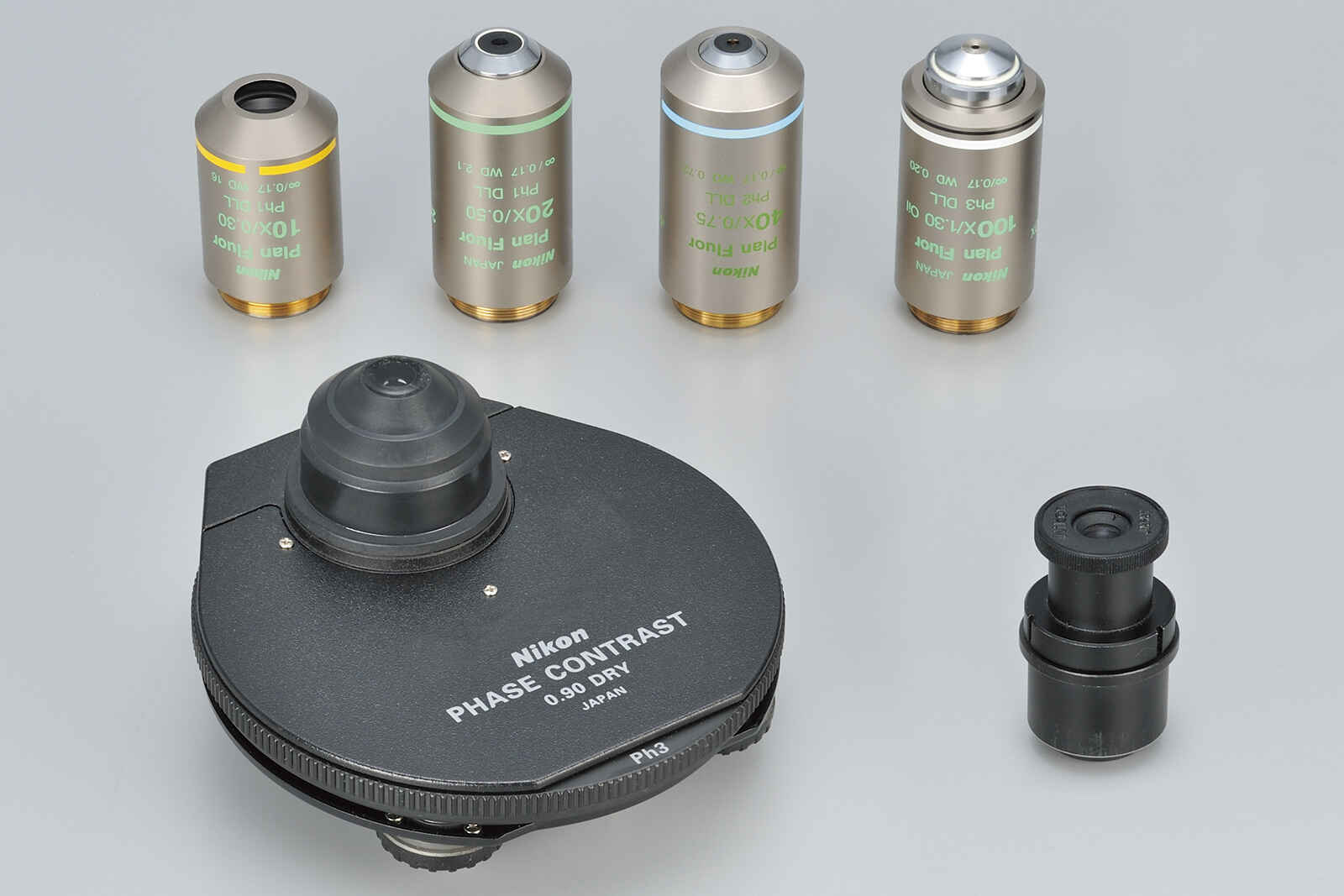
Accessories
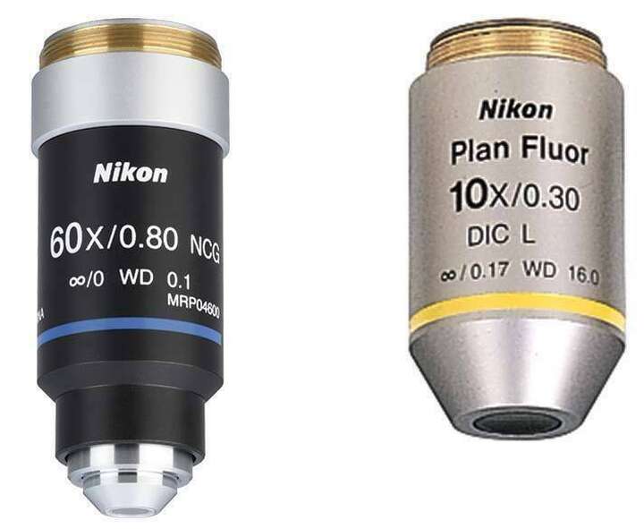
Objective lenses to meet diverse needs
We offer a wide range of objective lenses that are optimal for laboratory and observation work such as the Plan Fluor 10X with its long working distance of 16 mm, and the CFI Plan Apochromat Lambda Series featuring the excellent quality in optical performance.
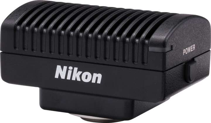
Microscope Digital Camera DS-Fi3
A 5.9 megapixel CMOS image sensor allows for the capture of images up to 2880 x 2048 pixels with color tones that faithfully represent the specimen.
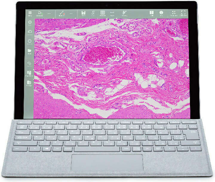
NIS-Elements L Imaging Software
By connecting the Digital Sight 1000 or DS-Fi3 to a tablet PC with NIS-Elements L installed, images of specimens under observation can be shared with other PCs via a network. The software also contains a variety of measurement and annotation functions.
Specifications
| Ci-E | Ci-L plus | Ci-S | |
|---|---|---|---|
| Optical system | CFI60 Infinity Optical System | ||
| Illumination | High luminescent White LED Illuminator | 6V30W Halogen Lamp Built-in ND4, ND8, NCB11 filters |
|
| Automatic intensity reproduction function | Light Intensity Management (LIM) feature, ECO mode*1, Sleep mode*2 | — | |
| Controls | Image capture button | ||
| Nosepiece rotating buttons Remote control pad |
— | ND filter IN/OUT switches | |
| Eyepieces (F.O.V. mm) |
Sleeve diameter Φ30mm
|
||
| Focusing | Coaxial Coarse/Fine focusing, Focusing stroke: 30 mm, Coarse: 9.33 mm/rotation, Fine: 0.1 mm/rotation, Fine movement scale 1μm, Coarse motion torque adjustable, Refocusing function | ||
| Tubes F.O.V. 22 mm (Eyepiece/Port) |
C-TB Binocular Tube C-TE2 Ergonomic Binocular Tube (Eyepiece: Port = 100:0, 50:50) via optional C-TEP2 DSC Port, C-TEP3 DSC Port C-0.55X Inclination angle: 10-30 degree, Extension: up to 40 mm |
||
| Tubes F.O.V. 25 mm (Eyepiece/Port) |
F-CTF Trinocular Tube F (Eyepiece: Port = 100:0, 0:100) TC-TT Trinocular Tube T (Eyepiece: Port = 100:0, 20:80, 0:100) |
||
| Nosepieces | Motorized Sextuple Nosepiece with Analyzer Slot (Within main body)
Switching between two objectives function*3 |
Exclusive Intelligent sextuple nosepiece (with Analyzer slot) | C-N6 ESD Sextuple Nosepiece ESD C-N6A Sextuple Nosepiece with Analyzer Slot |
| Stages | Cross travel 78 (X) × 54 (Y) mm, with vernier calibrations, stage handle height and torque adjustable for all stages
|
||
| Condensers (NA) Motorized |
CI-C-E Motorized Swing-out Condenser (0.9/0.22)
Focusing stroke: 27 mm |
— | |
| Condensers (NA) Manual |
Focusing stroke: 27 mm
|
||
| Observation methods*4 | Brightfield, Epi-fluorescence, Darkfield, Phase contrast, Simple polarizing, Sensitive color polarizing | ||
| Epi-fluorescence attachment | CI-FL-2 Epi-fluorescence Attachment (4 filter cubes mountable) D-FL-2 U-EPI Epi-fluorescence Attachment (6 filter cubes mountable, Terminator mechanism) |
||
| Epi-fluorescence light source | D-LEDI Fluorescence LED Illumination system C-HGFI/HGFIE HG Precentered Fiber Illuminator Intensilight (130W) |
||
| Power consumption | 13W (Brightfield configuration) | 5.0W(Brightfield configuration) | 38W (Brightfield configuration) |
| Weight (approx.) | 15.4 kg (Binocular standard set) | 13.4 kg (Binocular standard set) | 13.4 kg (Binocular standard set) |
*1 This is an energy-saving function that turns off the transmitted lighting and liquid crystal display to put the power consumption into a low power consumption state (Sleep mode) when there is no operation for a certain period of time.
*2 While the attached AC adapter is connected to the microscope, it is always energized, but it is in a state of standby with low power consumption.
*3 Settings with the remote control pad are required.
*4 Optional accessories are required for observations other than bright field.